
Page 94 - Libro 2
P. 94
74
PART 2 — CEREBROVASCULAR
RADIATION-INDUCED ARTERIAL INJURY
Radiation-induced arterial injury (RIAI) is caused by the use of therapeutic irradiation during treatment for various tumors. The treatment with radiation preferentially injures cancer cells with less injury to other tissues. However, there is a potential effect on blood vessels due to the presence of endothelial cells, which are sensitive to the radiation. Capillaries, ar- terioles, and venules are primarily involved, but it may also affect the carotid arteries in some patients. Injury to the vasa vasorum in the medial layer of the artery causes fibrosis. This and the repopulation of the endothelium may result in a narrowing of the lumen. Other risk factors, such as hypercholesterol- emia and hypertension, may add to the effect of the radiation, but not all patients develop these lesions.11
SIGNS AND SYMPTOMS
Patients will present with a history of radiation often several years prior to their examination. Often, these patients lack the typical risk factors for atherosclero- sis. An atypical location of an atherosclerotic steno- sis may tip the examiner to RIAI. In many patients, there is an absence of other atherosclerotic plaque in the carotids, which makes a single unusually located stenosis suspicious. TIA or CVA may be the effects of these lesions.
SONOGRAPHIC SCANNING TECHNIQUES
The distribution, extent of stenosis, and sonograph- ic characteristics of radiation-induced disease are different from the commonly encountered athero- sclerotic disease. The ultrasound examination must include thorough B-mode imaging of the common carotid arteries as there is a higher incidence of common carotid artery radiation-induced stenosis as compared to bifurcation and ICA disease. Trans- verse and longitudinal B-mode images should be taken to document the echogenicity of these lesions. As with all carotid examinations, spectral Doppler and color-flow imaging are used to assess the areas of stenosis.
Technical Considerations
A standard carotid duplex ultrasound examination is performed, although imaging may be difficult in some patients due to the changes in the tissue from the radiation. Poor echogenicity and hard, rather than supple neck tissue is common in postradiat- ed patients. Any surgery that may have altered the
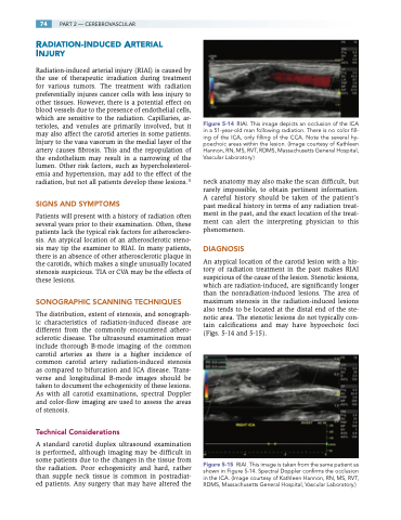
Figure 5-14 RIAI. This image depicts an occlusion of the ICA in a 51-year-old man following radiation. There is no color fill- ing of the ICA, only filling of the CCA. Note the several hy- poechoic areas within the lesion. (Image courtesy of Kathleen Hannon, RN, MS, RVT, RDMS, Massachusetts General Hospital, Vascular Laboratory.)
neck anatomy may also make the scan difficult, but rarely impossible, to obtain pertinent information. A careful history should be taken of the patient’s past medical history in terms of any radiation treat- ment in the past, and the exact location of the treat- ment can alert the interpreting physician to this phenomenon.
DIAGNOSIS
An atypical location of the carotid lesion with a his- tory of radiation treatment in the past makes RIAI suspicious of the cause of the lesion. Stenotic lesions, which are radiation-induced, are significantly longer than the nonradiation-induced lesions. The area of maximum stenosis in the radiation-induced lesions also tends to be located at the distal end of the ste- notic area. The stenotic lesions do not typically con- tain calcifications and may have hypoechoic foci (Figs. 5-14 and 5-15).
RIAI. This image is taken from the same patient as shown in Figure 5-14. Spectral Doppler confirms the occlusion in the ICA. (Image courtesy of Kathleen Hannon, RN, MS, RVT, RDMS, Massachusetts General Hospital, Vascular Laboratory.)
Figure 5-15
