
Page 96 - Libro 2
P. 96
76 PART 2 — CEREBROVASCULAR
disease, which is often eccentric and irregular. These patients will have abnormalities primarily related to the proximal common carotid arteries and subcla- vian arteries, sometimes with abnormal waveforms indicating proximal disease in the aorta or the ori- gins of the great vessels. If the aortic arch is affected proximally, bilateral changes in the common carotid waveforms will be noted, such as slow upstroke and lower than typical velocities bilaterally. If the aorta is stenosed in the area between the brachiocephalic and
the left common carotid, the left CCA and left subcla- vian artery waveforms may be turbulent, dampened, and display lower velocities than the right CCA and subclavian arteries.
An echolucent halo around the temporal artery is a strong indicator of temporal arteritis. Recent studies on the diagnostic value of the halo report sensitivity of 75% to 86% and specificity of 83% to 92% in pa- tients with temporal arteritis.13,14 Again, intermittent areas of focal velocity increases will be observed.
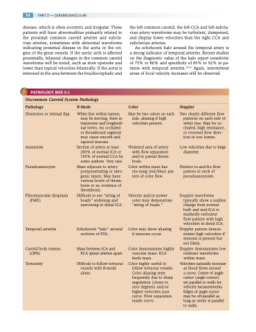
PATHOLOGY BOX 5-1
Uncommon Carotid System Pathology
Pathology B-Mode Color Doppler
Dissection or intimal flap
Aneurysm
Pseudoaneurysm
Fibromuscular dysplasia (FMD)
Temporal arteritis
Carotid body tumors (CBTs)
Tortuosity
White line within lumen; may be moving. Seen in transverse and longitudi- nal views. An occluded or thrombosed segment may cause smooth and tapered stenosis.
Section of artery at least 200% of normal ICA or 150% of normal CCA by some authors. Very rare.
Mass adjacent to artery postpenetrating or iatro- genic injury. May have various levels of throm- bosis or no evidence of thrombosis.
Difficult to see “string of beads” widening and narrowing in distal ICA.
Echolucent “halo” around sections of STA.
Mass between ICA and ECA splays arteries apart.
Difficult to follow tortuous vessels with B-mode alone.
May be two colors on each side, aliasing if high velocities present.
Widened area of artery with flow separation and/or partial throm- bosis.
Color within mass has yin-yang (red/blue) pat- tern of color flow.
Velocity and/or power color may demonstrate “string of beads.”
Color may show aliasing if stenoses occur.
Color demonstrates highly vascular mass. ECA feeds mass.
Color highly useful to follow tortuous vessels. Color aliasing seen frequently due to sharp angulation (closer to zero degrees) and/or higher velocities past curve. Flow separation inside curve.
Two clearly different flow patterns on each side of white line. May be oc- cluded, high resistance, or reversed flow direc- tion in one lumen.
Low velocities due to large diameter.
Distinct to-and-fro flow pattern in neck of pseudoaneurysm.
Doppler waveforms typically show a sudden change from normal bulb and mid-ICA to markedly turbulent flow pattern with high velocities in distal ICA.
Doppler pattern demon- strates high velocities if stenosis is present but not likely.
Doppler demonstrates low resistant waveforms within mass.
Velocities naturally increase as blood flows around
a curve. Center of angle cursor (angle correct)
set parallel to walls for velocity measurements. Edges of angle cursor may be off-parallel as long as center is parallel to walls.
