
Page 92 - Libro 2
P. 92
72
PART 2 — CEREBROVASCULAR
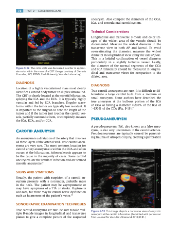
Figure 5-12 The color scale was decreased in order to appreci- ate color within the mass of a CBT. (Image courtesy of Damaris Gonzalez, RVT, RDMS, Rush University, Vascular Laboratory.)
DIAGNOSIS
Location of a highly vascularized mass most clearly identifies a carotid body tumor on duplex ultrasound. The CBT is clearly located at the carotid bifurcation, splaying the ICA and the ECA. It is typically highly vascular and fed by ECA branches. Doppler wave- forms within the tumor are typically low resistant. It is important to the surgeon to note the length of the tumor and if the tumor just touches the carotid ves- sels, partially surrounds them, or completely encases the ICA, ECA, and/or CCA.
CAROTID ANEURYSM
An aneurysm is a dilatation of the artery that involves all three layers of the arterial wall. True carotid aneu- rysms are very rare. The most common location for carotid artery aneurysms is within the CCA and often occurs at the bifurcation. Atherosclerosis appears to be the cause in the majority of cases. Some carotid aneurysms are the result of infection and are termed mycotic aneurysms.9
SIGNS AND SYMPTOMS
Usually, the patient with suspicion of a carotid an- eurysm presents with a nontender, pulsatile mass in the neck. The patient may be asymptomatic or may have symptoms of a TIA or stroke. Rupture is also rare, but there may be cranial nerve dysfunction such as hoarseness of the patient’s voice.10
SONOGRAPHIC EXAMINATION TECHNIQUES
True carotid aneurysms are rare. Be sure to take mul- tiple B-mode images in longitudinal and transverse planes to give a complete picture of the suspected
aneurysm. Also compare the diameters of the CCA, ICA, and contralateral carotid system.
Technical Considerations
Longitudinal and transverse B-mode and color im- ages of the widest area of the vessels should be documented. Measure the widest diameter in the transverse view in both AP and lateral. To avoid overestimating the diameter, measure the widest diameter in longitudinal view along the axis of flow. This is a helpful confirmation of vessel diameter particularly in a slightly tortuous vessel. Lastly, the diameter of the normal segments of the CCA and ICA bilaterally should be measured in longitu- dinal and transverse views for comparison to the dilated area.
DIAGNOSIS
True carotid aneurysms are rare. It is difficult to dif- ferentiate a large carotid bulb from a medium or small aneurysm. Some authors have described the true aneurysm at the bulbous portion of the ICA or CCA as having a diameter 200% of the ICA or 150% of the CCA (Fig. 5-13).9
PSEUDOANEURYSM
A pseudoaneurysm (PA), also known as a false aneu- rysm, is also very uncommon in the carotid arteries. Pseudoaneurysms are typically caused by penetrat- ing trauma or iatrogenic injury, creating a perforation
Figure 5-13 This image depicts a transverse view of a mycotic aneurysm at the carotid bifurcation. (Reprinted with permission from Journal for Vascular Ultrasound 2010;34:81.)
