
Page 91 - Libro 2
P. 91
5 — Uncommon Pathology of the Carotid System
71
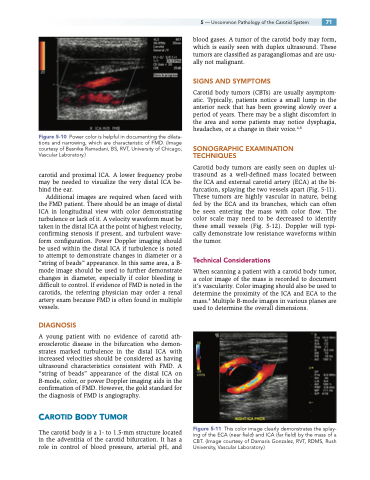
Figure 5-10 Power color is helpful in documenting the dilata- tions and narrowing, which are characteristic of FMD. (Image courtesy of Besnike Ramadani, BS, RVT, University of Chicago, Vascular Laboratory.)
carotid and proximal ICA. A lower frequency probe may be needed to visualize the very distal ICA be- hind the ear.
Additional images are required when faced with the FMD patient. There should be an image of distal ICA in longitudinal view with color demonstrating turbulence or lack of it. A velocity waveform must be taken in the distal ICA at the point of highest velocity, confirming stenosis if present, and turbulent wave- form configuration. Power Doppler imaging should be used within the distal ICA if turbulence is noted to attempt to demonstrate changes in diameter or a “string of beads” appearance. In this same area, a B- mode image should be used to further demonstrate changes in diameter, especially if color bleeding is difficult to control. If evidence of FMD is noted in the carotids, the referring physician may order a renal artery exam because FMD is often found in multiple vessels.
DIAGNOSIS
A young patient with no evidence of carotid ath- erosclerotic disease in the bifurcation who demon- strates marked turbulence in the distal ICA with increased velocities should be considered as having ultrasound characteristics consistent with FMD. A “string of beads” appearance of the distal ICA on B-mode, color, or power Doppler imaging aids in the confirmation of FMD. However, the gold standard for the diagnosis of FMD is angiography.
CAROTID BODY TUMOR
The carotid body is a 1- to 1.5-mm structure located in the adventitia of the carotid bifurcation. It has a role in control of blood pressure, arterial pH, and
blood gases. A tumor of the carotid body may form, which is easily seen with duplex ultrasound. These tumors are classified as paragangliomas and are usu- ally not malignant.
SIGNS AND SYMPTOMS
Carotid body tumors (CBTs) are usually asymptom- atic. Typically, patients notice a small lump in the anterior neck that has been growing slowly over a period of years. There may be a slight discomfort in the area and some patients may notice dysphagia, headaches, or a change in their voice.4,8
SONOGRAPHIC EXAMINATION TECHNIQUES
Carotid body tumors are easily seen on duplex ul- trasound as a well-defined mass located between the ICA and external carotid artery (ECA) at the bi- furcation, splaying the two vessels apart (Fig. 5-11). These tumors are highly vascular in nature, being fed by the ECA and its branches, which can often be seen entering the mass with color flow. The color scale may need to be decreased to identify these small vessels (Fig. 5-12). Doppler will typi- cally demonstrate low resistance waveforms within the tumor.
Technical Considerations
When scanning a patient with a carotid body tumor, a color image of the mass is recorded to document it’s vascularity. Color imaging should also be used to determine the proximity of the ICA and ECA to the mass.4 Multiple B-mode images in various planes are used to determine the overall dimensions.
Figure 5-11 This color image clearly demonstrates the splay- ing of the ECA (near field) and ICA (far field) by the mass of a CBT. (Image courtesy of Damaris Gonzalez, RVT, RDMS, Rush University, Vascular Laboratory.)
