
Page 90 - Libro 2
P. 90
70
PART 2 — CEREBROVASCULAR
stenosis in this case is typically smooth and tapered in nature, in the proximal/mid-CCA or mid-/distal ICA beyond the bifurcation. It is difficult to com- pletely differentiate between this situation and a long smooth atherosclerotic plaque; however, there will likely be more evidence of atherosclerosis in adjacent arteries in the latter case. Color changes are likely to be noticed in either the proximal and mid-CCA, or in the mid-/distal ICA beyond the bifurcation. An im- portant characteristic to identify the dissected artery is the distinctly different flow patterns in the true and false lumens by analysis of the Doppler waveforms.
FIBROMUSCULAR DYSPLASIA
Fibromuscular dysplasia (FMD) is a disease involv- ing abnormal growth of the arterial wall and may involve the intima, media, and/or adventitia. The media is the most common location for the abnor- mal growth of smooth muscle cells and fibrous tis- sue. The abnormal growth will sometimes cause narrowing of the arterial lumen in multiple sections with normal walls or slight aneurysmal dilatation in between the stenotic segments. This essentially causes a “string of beads” appearance of the artery on arteriography with larger and smaller diameters in sequence.
SIGNS AND SYMPTOMS
Fibromuscular dysplasia is primarily seen in young (25 to 50 years old) Caucasians, occurring in females three times more commonly than men.4–7 The most common location of disease is the renal arteries, and hypertension is frequently seen with renal involve- ment. The second most common vessel impacted is the internal carotid artery. Patients with carotid FMD often have no symptoms and can present with a cer- vical bruit. Embolization may occur and cause TIAs. In addition, approximately 30% of patients’ carotid FMDs may have cerebral aneurysms.
SONOGRAPHIC EXAMINATION TECHNIQUES
A young person who is referred to the vascular labo- ratory for a carotid duplex examination should be evaluated for evidence of FMD. Bilateral disease is typical. The typical “string of beads” appearance of the artery may not be clearly appreciated at first be- cause this disease primarily involves the distal ICA, where the vessel often courses deeply into the tis- sue, making detailed images difficult. The first sign of FMD during a duplex exam is most likely to be a sudden turbulence with high velocities in the distal
Figure 5-8 An internal carotid artery with FMD. In the area of FMD in the mid-/distal ICA, the Doppler spectral waveform demonstrates marked turbulence with spectral broadening that is clearly different than the normal waveform seen in the proximal ICA. (Image courtesy of Besnike Ramadani, BS, RVT, University of Chicago, Vascular Laboratory.)
ICA after the proximal arteries have shown no sign of atherosclerosis (Fig. 5-8). The sonographer may need to change to a lower frequency probe to vi- sualize more distally in the internal carotid artery (Fig. 5-9). Power Doppler may be helpful in visualiz- ing the characteristic beading in the artery, avoiding the distraction of aliasing from the marked turbu- lence (Fig. 5-10).
Technical Considerations
The distal ICA should be carefully examined with color, Doppler, and B-mode looking for a sudden tur- bulence distally after a normal appearing common
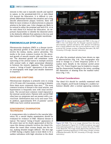
Figure 5-9 FMD. This is an image obtained from the same patient illustrated in Figure 5-8. A 3-MHz transducer was used and positioned posterior to the ear to obtain this view of FMD in the distal ICA. The image clearly depicts the turbulence and suggests a beading appearance of the artery. (Image courtesy of Besnike Ramadani, BS, RVT, University of Chicago, Vascular Laboratory.)
