
Page 88 - Libro 2
P. 88
68
PART 2 — CEREBROVASCULAR
SIGNS AND SYMPTOMS
A carotid artery dissection usually originates in the aorta and extends into the CCA. Some of these dis- sections that originate within the aortic arch may be associated with diseases such as Marfan’s syndrome or Ehlers-Danlos syndrome. However, it may also originate in the distal ICA and extend proximally or it may appear at the distal end of the carotid bifurca- tion during a duplex scan. These dissections can be either spontaneous or traumatic. Following carotid endarterectomy, an intimal flap may also form in the area of the endarterectomized segment.
A carotid dissection may be an unexpected finding in those patients where the dissection extends from the aorta because these dissections may cause no cerebral symptoms or pain. Although pain is not a typical symp- tom of patients with atherosclerosis, the patient with a dissection, particularly one that begins in the internal carotid artery, may present with pain in the head, face, or neck, or with or without hemispheric symptoms. Dissection is usually suspected when a young patient (typically 35 to 50 years old) presents with symptoms of stroke despite having no risk factors for atheroscle- rosis and particularly with a history of trauma. The trauma that may cause a dissection may not be evi- dent at first because it could be as subtle as a cough or turning the head or it could be a more obvious blunt trauma to the head or neck. Stretching of the artery may have occurred during these events, causing a tear in the arterial wall, or there may be an intimal weak- ness predisposing the individual to this consequence. With dissections that appear to be spontaneous, the primary risk factor is often hypertension.3–6
SONOGRAPHIC EXAMINATION TECHNIQUES
The first duplex ultrasound finding indicating that a dissection may be present is either an unusual color pattern in a section of an artery that otherwise shows no signs of atherosclerosis or the presence of a thin white line in the lumen that appears to flutter with each pulse. In B-mode, it is important to investigate the continued presence of the white line in both lon- gitudinal and transverse views and to be sure that it is not a refraction artifact from a nearby venous valve (Figs. 5-5 and 5-6). If the white structure can be visualized in multiple views, including anterior– posterior (AP) and lateral planes, it is much more likely to be a dissection rather than an artifact. If the media is also involved, the white structure may ap- pear thicker and may move less.
The flow patterns on each side of the dissected intima need to be investigated with Doppler, and each will be quite different (Fig. 5-7A–C). In the case where there is only one tear in the intima, blood
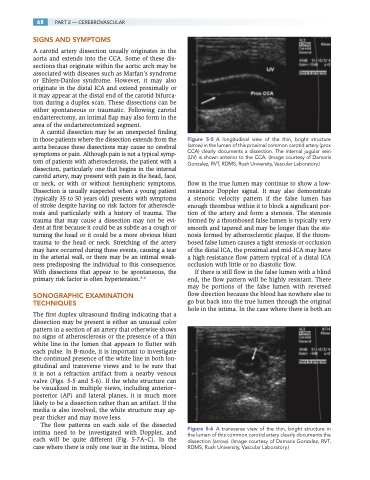
Figure 5-5 A longitudinal view of the thin, bright structure (arrow) in the lumen of this proximal common carotid artery (prox CCA) clearly documents a dissection. The internal jugular vein (IJV) is shown anterior to the CCA. (Image courtesy of Damaris Gonzalez, RVT, RDMS, Rush University, Vascular Laboratory.)
flow in the true lumen may continue to show a low- resistance Doppler signal. It may also demonstrate a stenotic velocity pattern if the false lumen has enough thrombus within it to block a significant por- tion of the artery and form a stenosis. The stenosis formed by a thrombosed false lumen is typically very smooth and tapered and may be longer than the ste- nosis formed by atherosclerotic plaque. If the throm- bosed false lumen causes a tight stenosis or occlusion of the distal ICA, the proximal and mid-ICA may have a high resistance flow pattern typical of a distal ICA occlusion with little or no diastolic flow.
If there is still flow in the false lumen with a blind end, the flow pattern will be highly resistant. There may be portions of the false lumen with reversed flow direction because the blood has nowhere else to go but back into the true lumen through the original hole in the intima. In the case where there is both an
Figure 5-6 A transverse view of the thin, bright structure in the lumen of this common carotid artery clearly documents the dissection (arrow). (Image courtesy of Damaris Gonzalez, RVT, RDMS, Rush University, Vascular Laboratory.)
