
Page 87 - Libro 2
P. 87
5 — Uncommon Pathology of the Carotid System
67
Figure 5-3 Appropriate angle correction techniques when sampling along a curved portion of the vessel.
lower velocities and demonstrate flow separation at or just distal to the curve. Some laboratories may choose to place the sample volume at the high- est point of velocity whether or not it is along the wall. Although this process will ensure that no high velocities will be missed, in the case of the curved vessel, a sample volume placed along the outer wall will possibly make the interpretation even more difficult as velocities measured here will be more likely to enter the abnormal range even in a normal artery.
Technical Considerations
A standard carotid duplex examination, as described in the preceding chapter, should be completed first. Additional transverse and longitudinal color im- ages of the tortuous carotid segments should be obtained. Scroll the sample volume through the tortuous segment or kink and document Doppler velocities pre- and post-curve or kink, setting the middle of the angle-correct cursor parallel to the walls of the vessel, as described previously. Trans- verse and longitudinal B-mode images should be used to identify any plaque along the curved seg- ment. A transverse B-mode image may be helpful in identifying the diameter of the artery at a tight kink. Some laboratories may document flow changes past a kink and/or symptoms while the patient turns his or her head.
DIAGNOSIS
High velocities naturally occur past a curve, making it difficult to apply strict velocity criteria on tortuous vessels. However, recognizing this normal flow phe- nomenon should be helpful to identify false-positive velocity increases when the arterial lumen appears normal. Unfortunately, there is no specific velocity criterion to apply to tortuous vessels due to the va- riety of angulations that may occur. Careful analysis of B-mode images with and without color in multiple
planes can be helpful as adjunct data to confirm that velocity changes around the curve are due to plaque formation or a significant kink rather than just a nor- mal flow response to a curve in the vessel. In the case of a significant stenosis, poststenotic turbulence will be present and will persist beyond the area in question.
DISSECTIONS/INTIMAL FLAPS
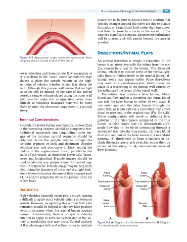
An arterial dissection is simply a separation of the layers of an artery, typically the intima from the me- dia, caused by a tear in the intima. The dissected intima, which may include some of the media, typi- cally flaps or flutters freely in the arterial lumen, al- though some may appear stable. Some dissections may result in a pseudoaneurysm, which forms be- cause of a weakening in the arterial wall caused by the splitting of the layers of the vessel wall.
The intimal tear creates a false lumen, where blood can flow and/or a thrombus can form. Blood can exit the false lumen in either of two ways. It can enter and exit the false lumen through the same tear, or it can exit via a secondary tear either distal or proximal to the original tear (Fig. 5-4A,B). Either configuration will result in differing flow patterns in the false lumen compared to the true lumen. A false lumen may (1) demonstrate ante- grade flow due to the blood continuing through a secondary tear into the true lumen, (2) have blood flow into and out of the false lumen in a to-and-fro pattern, (3) thrombose to form a stenosis or oc- clude the entire artery as it stretches across the true lumen of the artery, or (4) demonstrate reversed flow direction.
False lumen
True lumen
Dissected intima
False lumen ends
False lumen
True lumen
Dissected intima
Second tear
AB
Figure 5-4 A: Diagram of a blind-ended dissection. B: Diagram of a dissection with a secondary tear.
