
Page 86 - Libro 2
P. 86
66
PART 2 — CEREBROVASCULAR
very sharp angulation of the artery. As many as one- quarter of adults will have some degree of angula- tion within their internal carotid artery (ICA). Often, this is a bilateral finding.
SIGNS AND SYMPTOMS
Tortuous carotid arteries are usually asymptomatic, but a kinked artery may cause symptoms of stroke or transient ischemic attack (TIA), particularly upon turning the head. Tortuous carotid arteries may be congenital, affecting more women than men, but most patients with symptoms are of the older adult population.1 Frequently, a referring physician will mistake a very tortuous proximal common carotid (CCA) or brachiocephalic (innominate) artery for a carotid aneurysm because a large tortuous vessel that courses superficially may appear to be a very pulsatile mass upon palpation and/or upon being seen pulsing in the proximal neck. A duplex ultra- sound scan can easily differentiate the two.
SONOGRAPHIC EXAMINATION TECHNIQUES
Although tortuosity is not uncommon in the CCA, the ICA is actually more likely to be redundant. It is a challenge to follow these vessels in long and transverse views and takes considerable skill and experience to identify the course of the vessel as it changes planes and while it curves, loops, and some- times kinks (Fig. 5-1). Color is nearly essential to follow tortuous arteries and care must be taken not to accidentally move on to a branch that the tortuous artery may be crossing. Moving slowly up the neck
Figure 5-1 Color is a useful tool to appreciate the tortuous course of this distal ICA. (Image courtesy of Kimberly Gaydula, BS, RVT, University of Chicago, Vascular Laboratory.)
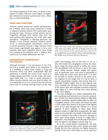
Figure 5-2 Color clearly demonstrates a looped internal ca- rotid artery with higher velocities (aliasing) along the outer walls of the vessel as the flow courses upward on the left side of the image and as it moves downward in the red segment in the middle of the image. (Image courtesy of Damaris Gonzalez, RVT, RDMS, Rush University, Vascular Laboratory.)
while concentrating only on the next 0.5 cm at a time will enable the sonographer to move the trans- ducer along the plane needed to follow the vessel. The sonographer will be forced to move the trans- ducer along unusual oblique angles and planes and, in transverse view, even move it proximally at times while the vessel turns upon itself. It is often not possible to obtain a picture of the entire tortu- ous segment in one image, but if it can be done, it creates a beautifully complex color picture (Fig. 5-2). Although Doppler angles close to zero degrees may occur causing color aliasing in the vertical segments of the artery, flow also naturally increases along the outer edges of a curve and may also cause aliasing in the color display.
The next challenge is to obtain interpretable Dop- pler waveforms and velocities on a tortuous carotid ar- tery.2 If there is no atherosclerotic disease or tight kink, Doppler waveforms should preferentially be taken on a straighter portion of the vessel, just before and after a curve, rather than directly at the point of the tightest curve. If you must measure the velocity on a curved segment of artery (perhaps in those cases where plaque is noted or a tight kink is suspected), the angle cursor is set so that the very middle of the angle-correct cursor is parallel to the walls of the artery (perhaps in those cases where plaque is noted or a tight kink is suspected), even though one or both ends of the cursor may not appear to be aligned correctly (Fig. 5-3)
As in any carotid duplex examination, the sam- ple volume should be kept center stream and small despite the fact that velocities are higher along the outside of the curve. The inside of a curve may have
