
Page 4 - Articulo: Musculoesqueletal ultrasound
P. 4
Musculoskeletal Ultrasound: A Primer for Primary Care
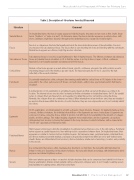
Table 2. Description of Structures Seen by Ultrasound
Structure
Comment
Skeletal Muscle
On longitudinal views, the muscle septae appear as bright/echogenic structures and are seen as thin, bright, linear bands (“feathers” or “veins on a leaf”). On transverse views, the muscle bundles appear as speckled echoes, with short, curvilinear, bright lines dispersed throughout the darker/hypoechoic background (“starry night”).
Fascia
Fascia is a collagenous structure that usually surrounds the musculotendinous areas of the extremities. Fascia is encompassed by subcutaneous tissue. The fascia often is seen inserting into bone and blending with the periosteum. Normal fascia appears as a fibrous, bright hyperechoic structure.
Subcutaneous Tissue
Subcutaneous tissue is isoechoic (equal brightness) to that of skeletal muscle. The difference between subcutaneous tissue and skeletal muscle visualized on US is that the septae do not lay in lines or layers. A thick, continuous hyperechoic band usually separates subcutaneous fat from muscle.
Cortical Bone
Normal cortical bone appears as a well-defined, linear, smooth, continuous echogenic line with posterior acoustic shadowing (image beyond the interface appears black). The hyperechogenicity of bone is caused by the high reflectivity of the acoustic interface.
Periosteum
Occasionally visualized as a thin, echogenic line running parallel with the cortical bone on US. Injuries to the bone— especially to the cortex, periosseous soft tissues, and periosteum—will produce a periosteal reaction that may be visualized.
Tendons
A normal tendon on US examination is a bright/echogenic linear band that can vary in thickness according to its location. The internal echoes are described as having a fibrillar echotexture on longitudinal views. On US, the parallel series of collagen fibers are hyperechoic and separated by darker/hypoechoic surrounding connective tissue. Normally, the collagen fibers are continuous and intact. When interruptions in tendon fibers exist, they are visualized as anechoic/black areas within the tendon. As solid structures, they are noncompressible and do not normally exhibit blood flow.
Ligaments
On US examination, a normal ligament is a bright, echogenic, linear structure. However, for ligaments having a more, compact, fibrillar echotexture, the individual strands/fibers of the ligaments are more closely aligned. Ligaments are composed of dense connective tissue, similar to tendons, but with much more variability in the amounts of collagen, elastin, and fibrocartilage. This makes imaging a ligament more variable than a tendon. Ligaments can easily be distinguished from tendons by tracing the ligament to the bony structures to which it attaches, with a characteristic “broom-end” appearance in transverse views.
Peripheral Nerves
High-frequency transducers allow the visualization of peripheral nerves that pass close to the skin surface. Peripheral nerves appear as parallel hyperechoic lines with hypoechoic separations between them. On longitudinal views, their appearance is similar to tendons but less bright/echogenic. On transverse views, peripheral nerves, individual fibers, and fibrous matrix present with multiple, punctuate echogenicities (bright dots) within an ovoid, well-defined nerve sheath. Nerves are differentiated from tendons by their echotexture, relative lack of anisotropy, location, and proximity to the vessels.
Bursae
In a normal joint, the bursa is a thin, black/anechoic line that is <2 mm thick. The bursa fills with fluid when it is irritated or infected. Depending on the extent of effusion, the bursa will distend and enlarge, with inflammatory debris expressed as internal brightness echoes.
Vessels
Veins and arteries appear as hypo- or anechoic tubular structures that can be compressed and exhibit blood flow on Doppler examination. Arteries remain pulsatile during compression, whereas veins do not. Usually, localizing vessels may facilitate in localizing nerves, which lie beside them.
November 2012 | Practical Pain Management 57
