
Page 5 - Articulo: Musculoesqueletal ultrasound
P. 5
Musculoskeletal Ultrasound: A Primer for Primary Care
A
Attenuation results in reduction of the acoustic energy and increases as a function of depth and frequency. These scanning skills require dedi- cation, training, and many hours of practice to master in the clinic.
US is very useful in diagnosing damages or injuries to structures. Table 3 reviews common injuries seen on US.1,11
Therapeutic Applications
In MSK US
The use of ultrasonography in inter- ventional MSK radiology is well established and is used primarily to guide needle placement for injec- tions, aspirations, and biopsies.12 The choice of US transducer is critical, with high-frequency (7-12 MHz) lin- ear array transducers being used most frequently. For deeper structures, such as hips, and larger patients, lower frequency curvilinear probes (4-9 MHz) be required, although they may be prone to anisotropic artifact. Regardless of the probe selected, a complete sonographic examination (including Doppler exam) of the proposed area should be conducted to determine critical structures such as nerves and vessels. This allows the determination of needle trajectory and avoidance of areas of potential infection.
Most interventional US procedures are performed with a “free-hand tech- nique,” which allows direct, dynamic visualization of the needle tip. After planning the safest route of needle access, a line parallel to the long axis of the probe face can be drawn on the skin. The patient’s skin and trans- ducer then are sterilized and draped. The needle is directed toward the intended target under vigilant obser- vation with the long axis of the needle and in line with the long axis of the transducer face.
Strategies to discriminate the needle
A
B
C
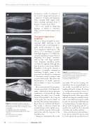
Figure 1. Suprapatellar aspiration. Ultrasound image prior to aspiration allowed determination of no internal debris in the often-recurring site of fluid accumulation.
A, longitudinal views of the suprapatellar pouch/bursa demonstrate a large anechoic fluid collection;
B, needle insertion for aspiration;
C, post aspiration view.
B
Figure 2. Supraspinatus injection.
A, longitudinal probe placement on the anterior/ lateral shoulder reveals a nearly full-thickness tear of the supraspinatus tendon;
B, ultrasound-guided injection.
tip under US involve keeping the transducer face as perpendicular to the needle as possible by heel-toe angling and probe rocking. By doing so, reverberation artifact posterior to the needle is seen and aids in high- lighting the needle. Other approaches include sweeping the transducer from side to side while moving the needle in and out; injecting a small amount of local anesthetic to localize the nee- dle tip; and rotating the probe 90° to examine the needle in short axis and determine the needle’s pathway.
Intra-articular interventional injec- tions incorporating US may be used for joint aspirations (eg, detection of
58
Practical Pain Management
|
November 2012
