
Page 7 - Articulo: Musculoesqueletal ultrasound
P. 7
Musculoskeletal Ultrasound: A Primer for Primary Care
An endurance sports enthusiast, Dr. Pinzon developed chronic right heel pain over the plantar fascia/heel region that has lasted 9 months (Figure). He developed right plantar fascia tendonosis and was treated with various conserva- tive treatments including: plantar fascia stretching, gel-heel pads, custom-molded orthotics, physi- cal therapy heel mobilization/ myofascial release techniques, top- ical analgesics, and heel counter in athletic footwear. After some failed conservative treatment options, he chose to have a musculoskel- etal ultrasound (MSK US)-trained specialist pursue sonographic- guided local corticosteroid injec- tion using a posteriomedial approach beneath and within the fascial substance, enabling infiltra- tion of a corticosteroid/anesthetic
solution. The solution of 20 mg of methylprednisolone and 0.5% marcaine was injected into the thickest part of the plantar fascia from its attachment to the calca- neous, without significant resis- tance noted. Subsequently cold/ ice treatment was applied and avoidance of significant weight- bearing was ordered for 2 days
after the procedure. Four weeks post-injection procedure and after continued stretching, topi- cal analgesics, and orthotics, Dr. Pinzon noted >90% improvement including dramatic improvement in his chronic right heel pain. Dr. Pinzon now is able to participate in some of his endurance sporting endeavors.13
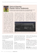
Clinical Vignette:
Plantar Fascia Tendonosis
Drawing from his own personal experience, the author presents a case of chronic right heel pain treated by ultrasound-guided therapy.
Figure. Plantar fascia foot comparison: affected and unaffected plantar fascia tendonitis.
probe oriented axially is preferred. With the femoral head and acetabular rim in view, the needle is introduced from an anterolateral approach.
Knee Joint
For distended knee joints with effu- sions, the suprapatellar bursa, the best access is usually with the patient lying supine with the knee flexed slightly. The probe is held parallel to the quad- riceps tendon and slid medially or lat- erally until the quadriceps fibers dis- appear and the needle is directed into the bursa. For knee joints without effu- sions, the medial patellofemoral facet
is the best target, with the probe in the axial plane of the patella and medial femoral condyle visible. The probe is turned 90° and oriented along the joint line and the needle is then introduced either inferiorly or superiorly into the joint.
Ankle Joint
With the patient lying in supine posi- tion, the anterior tibiotalar joint is examined in a sagittal plane. The exam- iner may perform plantarflexion or dor- siflexion maneuvers to identify the talus movements across the tibia. The dorsa- lis pedis artery and extensor tendons
should be avoided. The needle entry into the joint is in a sagittal plane using an inferior approach.
Summary
The integration of diagnostic and inter- ventional MSK US into clinical practice is a welcome alternative to procedures that might otherwise be performed under fluoroscopic or computed tomo- graphic guidance in the fields of radiol- ogy, physiatry, and anesthesia.
Authors’ Bios: Elmer G. Pinzon, MD, MPH, FABPMR, is the president, medi- cal director, and owner of University
60
Practical Pain Management | November 2012
