
Page 6 - Articulo: Musculoesqueletal ultrasound
P. 6
Musculoskeletal Ultrasound: A Primer for Primary Care
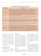
Table 3. Description of Common Injuries Seen on Ultrasound
Structural Injury
Description
Tendon Injuries
Tendonosis manifests as tendon enlargement, hypoechogenicity, and an increase in interfibrillar distance— primarily due to intratendinous edema. Partial-thickness tears present as additional findings of focal regions of anechogenicity accompanied by loss of the normal fibrillar pattern, but tendon continuity is maintained. High-grade, partial-thickness tearing is imaged as tendon thinning due to tendon substance loss. Full-thickness tearing is seen as tendon gaps occurring in conjunction with tendonosis-related changes. Tenosynovitis may appear either as simple anechoic with easily displaceable fluid surrounding the tendon or complex fluid with mixed echogenicity. Complex fluid seen on imaging within the tendon sheath should be diagnostically aspirated if infection is suspected.
Ligament Injuries
Low-grade injuries are imaged as enlarged, hypoechoic ligaments with normal echotexture, while partial- and full-thickness tears reveal fibrous disruption. Stress testing may be able to differentiate between partial versus complete tears and assess joint stability, as in the case of tendon pathology.
Nerve Injuries
Similar to tendons and ligaments, affected nerves reveal regional swelling, diffuse hypoechogenicity, and loss of fascicular pattern. A “notch sign” is a reflection of entrapment sites, which are localized by evaluating swelling proximal to the entrapment site and a focal narrowing at that site.
Muscle Injuries
Low-grade muscle strains exhibit subtle regions of hypoechogenicity accompanied by reduction in the normal pennate echotexture, making the affected area look “washed out.” High-grade contusions and injuries reveal variability in frank fiber disruption and heterogeneous fluid, as seen in hematomas.
Bone and Joint Disorders
Periostitis or stress fracture is seen with irregularities in the smooth, superficial surface of bone. Ultrasound is
very sensitive in the detection of joint effusions. Joint effusions are anechoic, compressible, and devoid of Doppler flow. Complex, heterogeneous-appearing fluid may be indicative of infection for which aspiration is recommended. Synovitis appears as noncompressible, echogenic tissue within a joint and hyperemia on Doppler. Periarticular erosions, crystal-related deposits, and gouty tophi also may be seen in the joint evaluation. Enlarged bursae contain simple anechoic fluid but, similar to joint effusions, may contain complex fluid. Periarticular and peritendinous ganglia may be present as multilobulated, anechoic noncompressible structures devoid of blood flow.
crystal arthropathy or septic arthri- tis; Figure 1) or therapeutic intra- articular injections with corticoste- roids or viscosupplementation (eg, treatment of joint arthritis; Figure 2). Diagnostic injections using short- and long-acting anesthetics can determine the patient’s symp- tom improvements with long-acting agents. Most hip and shoulder joints may accept up to 10 mL, but small joints of the hands and feet may only accept 1 to 2 mL.
Potential US-guided
Routes of Access
Some of the most promising routes of access to the most commonly injected joints under US guidance are presented here.12
Shoulder Joint
The patient is best positioned in a seated or lateral decubitus position. The patient’s hand is positioned rest- ing on the opposite shoulder, and the key landmarks of the triangular-shaped posterior labrum, humeral head, and joint capsule are identified. The gleno- humeral joint is best accessed from the posterior rather than anterior approach. The needle is introduced laterally in the axial plane and advanced medially, with the needle target between the posterior aspect of the humeral head and poste- rior labrum.
Elbow Joint
The patient is best positioned in a seated or supine position with elbow flexed and arm across the chest. The probe is
positioned along the posterior elbow and oriented sagittally with the triceps tendon longitudinally placed. The needle is introduced superiorly, passing beside the triceps tendon and through the posterior fat pad to enter the joint space. Key landmarks are the olecranon fossa of the humerus, posterior fat pad, and the olecranon.
Hip Joint
The patient lies supine, and the joint is accessed anteriorly. With joint effusions or larger patients, the optimal approach is with the probe aligned along the long access of the femoral neck. The needle is introduced from the inferior approach, passing through the joint capsule to rest on the subcapital femur. In thin- ner patients, easier access with the US
November 2012 |
Practical Pain Management 59
