
Page 105 - Libro vascular I
P. 105
Chap-08.qxd 1~9~04 16:41 Page 96
96
PERIPHERAL VASCULAR ULTRASOUND
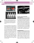
CCA
A: Color image showing a narrowed proximal ICA. B: Doppler recording obtained from within
the narrowing, demonstrating a significant velocity increase (peak systolic velocity 500 cm/s, end diastolic velocity 300 cm/s) with increased spectral broadening, suggesting a significant stenosis (80% diameter reduction).
greatest narrowing within a diseased segment of a vessel, which should then be investigated with the spectral Doppler (Fig. 8.17B). High-velocity jets may be seen within and just beyond a stenosis and the path of the flow may no longer be parallel to the vessel wall. In this case, the color image allows more accurate angle correction for velocity meas- urements. The complete absence of color Doppler filling within a vessel could indicate that the vessel is occluded, but this should be confirmed by opti- mizing the color controls for detection of low- velocity flow to rule out the presence of a very tight stenosis somewhere along the vessel. The apparent lack of color filling within the CCA or ICA during diastole may indicate high-resistance flow due to an occlusion or tight stenosis distally. Spectral Doppler should be used to confirm the absence of diastolic flow, and both color and spec- tral Doppler should be used to investigate the dis- tal vessels carefully.
ICA
A
CCA
Vein
ICA
Figure 8.18
A long-standing ICA occlusion.
SPECTRAL DOPPLER WAVEFORMS
Normal appearance
Spectral Doppler recordings obtained from the ECA show a high-resistance flow pattern with a pulsatile waveform shape and low diastolic flow (Fig. 8.9C) compared with the low-resistance waveform shape seen in the ICA (Fig. 8.9B). The normal CCA wave- form (Fig. 8.9A) has a shape somewhere between that of the ICA and the ECA. The peak systolic velocities seen in the carotid arteries depend on the relative size of the vessel but are typically less than 110 cm/s in the normal ICA. The flow profiles in the normal bifurcation seen in color flow imaging (see Fig. 5.12) will affect the spectral Doppler waveform shapes detected in the ICA origin, which may appear disturbed or demonstrate areas of reverse flow. Distal to the bifurcation, the wave- form shapes should no longer appear disturbed.
Abnormal appearance
The presence of a narrowing within the carotid arteries will lead to an increase in the velocity of the blood across the stenosis, and this can be measured using spectral Doppler. Significant changes in the velocity within and just beyond a stenosis will be detected once the vessel is narrowed by a 50% reduction in diameter. The increase in velocity is related to the degree of narrowing (see Ch. 5). These velocity changes can be used to grade the degree of narrowing. The Doppler waveforms obtained within or just beyond a significant stenosis
B
Figure 8.17
