
Page 106 - Libro vascular I
P. 106
Chap-08.qxd 1~9~04 16:41 Page 97
ULTRASOUND ASSESSMENT OF THE EXTRACRANIAL CEREBRAL CIRCULATION
97
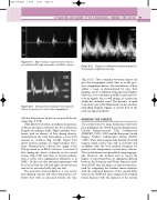
Figure 8.19 High-resistance waveform detected in a non-diseased ICA origin proximal to an MCA occlusion.
Figure 8.20 CCA waveform showing reverse diastolic flow in the presence of aortic valve regurgitation.
will also demonstrate an increase in spectral broad- ening (Fig. 8.17B).
Unusually low velocities can indicate the presence of disease proximal or distal to the site at which the Doppler recording is made. High-resistance wave- forms, with an absence of flow during diastole, obtained from the CCA may indicate a severe ICA stenosis or occlusion (Fig. 8.10B). Figure 8.19 shows another example of a high-resistance wave- form, obtained from a disease-free origin of the ICA proximal to an MCA occlusion. A reversal of flow during the whole of diastole in the carotid arteries (Fig. 8.20) may relate to a heart problem, such as aortic valve regurgitation (Malaterre et al 2001). In this case this abnormal appearance will be seen in both the left and right carotid arteries and not be associated with only one side.
The waveform detected distal to a very severe, flow-limiting stenosis will often demonstrate tur- bulent flow with an increased systolic rise time
Figure 8.21 Doppler recording demonstrating turbulent flow beyond a significant stenosis.
(Fig. 8.21). These untypical waveform shapes can give the sonographer useful clues as to the pres- ence of significant disease. The total absence of flow within a vessel, as demonstrated by color flow imaging, can be confirmed using spectral Doppler. However, it is sometimes possible to pick up low- velocity signals, due to wall thump, at a point just within the occluded vessel. The presence of small veins in the area of the bifurcation can also produce misleading Doppler signals, as venous flow in the neck can appear pulsatile.
GRADING THE DISEASE
The results from two large multicenter trials have been published, the North American Symptomatic Carotid Endarterectomy Trial Collaborators (NASCET) (1991, 1998) and the European Carotid Surgery Trialists’ Collaborative Group (ECST) (1998). These trials compared the benefits of carotid surgery, which carries some risk of mortality and morbidity, with the best medical treatment for patients with symptomatic carotid artery disease. The carotid disease was quantified using angiogra- phy. However, the method used to report the degree of narrowing from an angiogram differed between the European and North American trials. In the ECST trial, the degree of stenosis was meas- ured by comparing the residual lumen diameter with the estimated diameter of the carotid bulb, whereas the NASCET trial compared the residual lumen diameter with the diameter of the normal
