
Page 107 - Libro vascular I
P. 107
Chap-08.qxd 1~9~04 16:41 Page 98
98
PERIPHERAL VASCULAR ULTRASOUND
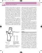
distal ICA, as shown in Figure 8.22. Using these two different methods can lead to significant dif- ferences in grading the disease. For example, the narrowing in Figure 8.22 would be reported as a 70% diameter reduction by the European method but as only a 50% reduction by the North American method. Figure 8.22 also gives the approximate equivalent degree of stenosis, measured (from the same stenoses) using the different methods employed by the NASCET and ECST trials. It has been proposed that a better method of measure- ment may be to compare the residual lumen with the diameter of the distal CCA.
The difference in methods used to grade the degree of narrowing has made the comparison of the results from the two trials complicated. The ECST study showed that surgery reduced the risk of stroke in patients with ECST70–99% stenosis. However the NASCET reported similar results for patients with NASCET70–99%, which is equivalent to a ECST80–99% stenosis. Taking the results of both trials together Rothwell (2000) concluded that carotid
endarterectomy reduces the overall risk of stroke in patients with a recently symptomatic ECST70–99% stenosis (NASCET50–99%). Rothwell states that using these criteria, it would be necessary to operate on 8 to 10 patients to prevent one stroke over the next 3 years. Another trial studying patients with signifi- cant (60–99%) asymptomatic stenosis, the Asympto- matic Carotid Atherosclerosis Study (ACAS) (1995), showed limited benefit of surgery in this group. Future developments in patient selection may enable the group of patients at high risk of stroke to be more closely targeted. The fact that the symptoms are likely to relate to embolic rather than hemo- dynamic phenomena means that there is still some clinical debate as to whether plaque type and volume, along with the degree of vessel narrowing, are criti- cal factors in the cause of stroke.
Angioplasty and stenting can also be used to treat carotid artery stenosis, as an alternative to endarterectomy, but the risks involved still make this a controversial method requiring randomized con- trolled trials comparing the two methods of treat- ment (Rothwell 2000). Stents are expandable mesh tubes that can be used to keep a diseased vessel patent. Although stent placement does not involve a general anesthetic, unlike carotid endarterec- tomy, there is a potential risk of stroke during the procedure.
Imaging
Angiographic grading of carotid artery disease, as with other arterial disease, is described in terms of diameter reduction. Therefore, ultrasound grading of stenoses is also typically described in terms of diameter reduction, although the use of area reduction would seem more appropriate, especially in the presence of eccentric disease. Table 8.1 gives the percentage area reduction associated with a given percentage diameter reduction, assuming a symmetrical lumen reduction; however, these val- ues are not correct in the presence of eccentric dis- ease. B-mode imaging is the most appropriate method to evaluate the degree of narrowing, if the degree of lumen diameter reduction is less than 50%. However, if disease is eccentric, it is possible to overestimate the degree of narrowing if the atheroma lies on the anterior or posterior wall when imaged longitudinally. It is equally possible to underestimate the degree of narrowing on a
ICA
ECA
NASCET A B A
ECST CB C
CCA
Estimated position of vessel wall
NASCET ECST 30 65 40 70 50 75 60 80 70 85 80 91 90 97
Approximate equivalent degree of ICA stenosis according to NASCET and ECST measurement methods
Figure 8.22 The NASCET and ECST trials used different methods of reporting the degree of narrowing seen on carotid angiograms. (After Donnan et al 1998, with permission.)
B
C
A
