
Page 108 - Libro vascular I
P. 108
Chap-08.qxd 1~9~04 16:41 Page 99
longitudinal image if the plaque is situated on the lateral walls (Fig. 8.23). Therefore, the diseased vessel should be visualized in transverse section first, in order to select the optimal longitudinal imaging plane, although this is obviously limited by the range of longitudinal scan planes available.
The percentage diameter reduction can be esti- mated from diameter measurements as follows:
Color flow imaging can help in identifying any lumen reduction. It is possible to obtain a color flow image in longitudinal and transverse section and this may help in estimating the degree of
ULTRASOUND ASSESSMENT OF THE EXTRACRANIAL CEREBRAL CIRCULATION
99
Table 8.1 Relationship between diameter reduction and cross-sectional area reduction assuming a concentric stenosis
Diameter reduction (%)
30 50 70
Cross-sectional area reduction (%)
50 75 90
% diameter reduction
1
diameter of patent lumen
total diameter 100
of vessel
1
2
5
5
1
3
D
6 26
A
B
C
3
4
E
F
7
7
4
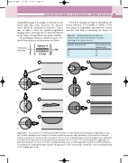
Figure 8.23
It is possible to overestimate and underestimate eccentric disease when imaging in longitudinal section. The schematic diagrams show examples of disease imaged in transverse and longitudinal section from the numbered transducer positions. A: An area of atheroma may not be seen in one longitudinal plane (1) and may appear more significant in another (2). B: Atheroma on the lateral walls may protrude into the centre of the vessel and give the appearance of atheroma floating in the vessel. C, D: These longitudinal images give a similar appearance despite very different degrees of narrowing. The longitudinal image may give the appearance of the vessel being occluded (E) or stenosed (F) depending on the imaging plane used.
