
Page 110 - Libro vascular I
P. 110
Chap-08.qxd 1~9~04 16:41 Page 101
ULTRASOUND ASSESSMENT OF THE EXTRACRANIAL CEREBRAL CIRCULATION
101
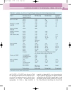
Table 8.2 Summary of a selection of reported Doppler ultrasound criteria for diagnosing stenosis
Author
Bluth et al (1988)
Robinson et al (1988) (NASCET)
Hunik et al (1993) (NASCET)
Fraught et al (1994) (NASCET)
Sidhu & Allan (1997)
Filis et al (2002) (NASCET)
Staikov et al (2002)
Grant et al (2003) (NASCET)
Percentage stenosis diameter reduction
40–59
60–79
80–99
50 50 70
70–99
50–69 70–99
50–59
60–69
70–79
80–95
96–99
100
50 50–59 60–69 70–79 80–89 90–99 Occlusion
70–99 (NASCET) 70–99 (ECST & CC†) 80–99 (ECST)
50
50–69
70 but less than near occlusion Near occlusion
ICA PSV (cm/s)
130 130 250
150 150 225
230 130
130 130
130 230 230 ‘String flow’ ‘No flow’
ICA EDV (cm/s)
40* 40* 100*
50 50 75
100 100
40 40–110 110–140 140
ICA PSV to CCA PSV ratio
1.8 1.8 3.7
2 2 3
3.2 3.2–4 4 4
Total occlusion *Peak diastolic velocity; †common carotid method.
220 190 215
125 125–230 230
High, low or undetectable Undetectable
80 65 90
40 40–100 100
Variable
Not applicable
150
150–200
200–250
250–330
330–400
400
‘No flow detected at ICA by PW/CDI using sensitive scale settings. Unilateral blunted CCA flow’
50 50–70 70–90 90–130 130–180 180
1.8 2.2 2.2–2.8 2.8–3.8 3.8–5 5
2.0 2.0–4.0 4.0
Variable
Not applicable
the ICA PSV to CCA EDV ratio, shown in Table 8.3. An important factor that may affect the crite- ria selected for grading stenoses is whether ultra- sound is to be used as a screening test before angiography, for which a high sensitivity is
required (see Appendix B), or to select patients for surger y, without angiography, for which sensitivity and specificity should both be equally high, to keep the number of false-positive results as low as possi- ble. The publications describing the criteria listed
