
Page 112 - Libro vascular I
P. 112
Chap-08.qxd 1~9~04 16:41 Page 103
imaged in a different plane (Fig. 8.24C) the flow can be seen within the vessel. Although the vessel lumen looks to be only slightly narrowed on the color image, the presence of a velocity increase, demon- strated by aliasing, should alert the sonographer to the possible presence of a more significant narrowing. Spectral Doppler velocity measurement (Fig. 8.24D) gives a PSV of 200 cm/s and an EDV of 75 cm/s in the ICA. Using the velocity criteria of Filis et al (2002) (Table 8.2) these velocity measurements would indicate a narrowing of 60–69% (NASCET) diameter reduction. By using the appearance of both B-mode and color images in transverse and a variety of longitudinal imaging planes, along with velocity measurements, the sonographer is able to estimate the degree of narrowing. There will, however, be sit- uations in which imaging and velocity measurement are limited and the sonographer is unable to make a judgment on the severity of the disease, and this should be made clear in the scan report.
NORMAL AND ABNORMAL APPEARANCES
OF VERTEBRAL ARTERY FLOW
The vertebral artery and vein can be seen between the vertebral processes. The vein normally lies
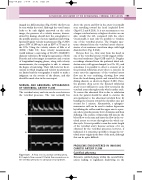
A
B
C
Figure 8.25 A: Flow seen in a normal vertebral artery. B: Complete flow reversal. C: Partial flow reversal seen in the vertebral artery due to subclavian steal syndrome.
ULTRASOUND ASSESSMENT OF THE EXTRACRANIAL CEREBRAL CIRCULATION
103
above the artery, and flow in the artery is normally seen travelling toward the head (cephaled flow) (Figs 8.12 and 8.25A). It is not uncommon to see a larger vertebral artery or higher velocities on one side, usually the left, compared with the other. Occasionally, it may only be possible to visualize one of the vertebral arteries. The Doppler spec- trum obtained from the vertebral artery demon- strates a low-resistance waveform shape with high diastolic flow (Fig. 8.25A).
Reverse flow (i.e., flow away from the head) in one of the vertebral arteries would suggest subcla- vian steal syndrome (Figs 8.25B and 8.3). Doppler recordings obtained from the ipsilateral distal sub- clavian artery will appear damped (see Ch. 10), and sometimes it is possible to detect a stenotic jet in the proximal subclavian artery due to a stenosis. In some cases the appearance of the vertebral artery flow can be very confusing, showing flow away from the head during systole and toward the head during diastole, as shown in Figure 8.25C. Here, the pressure drop across the diseased subclavian artery is not sufficient to cause flow reversal in the vertebral artery throughout the whole cardiac cycle. To ensure this abnormal flow is due to subclavian steal, the patient should be asked to exercise the arm ipsilateral to the abnormal vertebral flow, by bending the forearm toward the shoulder once per second for 1 minute. Alternatively, a sphygmo- manometer cuff can be used to induce hyperemia by inflating the cuff around the upper arm to a pres- sure above systolic pressure for 2–3 minutes and then deflating. The exercise or hyperemia will increase the blood flow to the arm and cause the flow in the ver- tebral artery to reverse throughout the whole car- diac cycle. It is not possible to scan the entire length of the vertebral artery because sections of it are obscured by the vertebral processes; however, if indicated, it is sometimes possible to image the ver- tebral artery origins in the base of the neck, although this can be quite difficult.
PROBLEMS ENCOUNTERED IN IMAGING CAROTID ARTERY FLOW
Calcified atheroma
Extensive calcified plaque within the carotid bifur- cation leading to significant shadowing on the
