
Page 113 - Libro vascular I
P. 113
Chap-08.qxd 1~9~04 16:42 Page 104
104
PERIPHERAL VASCULAR ULTRASOUND
B
CCA
Vein
ICA
A
ICA
CCA
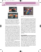
Figure 8.27
Color flow image of a tortuous ICA.
ICA
CCA
waveform beyond it cannot be used to indicate the absence of any significant narrowing as normal flow can be established within a short distance dis- tal to a stenosis. If an abnormal waveform is detected beyond the calcification, the presence of a signifi- cant stenosis can be more confidently predicted. High peak systolic and end diastolic velocities (Fig. 8.17B) produced by a jet extending beyond a stenosis, post-stenotic flow turbulence (Fig. 8.21) or low-velocity, damped flow would all suggest the presence of a significant stenosis. If any doubt about the presence or absence of significant disease remains at the end of the examination, the sono- grapher should make this clear in the report, as angiography may be required to clarify the degree of narrowing. In cases of less severe calcification, the sonographer may be able to overcome poor imaging by viewing the vessels in a different plane (Fig. 8.24).
Vessel tortuosity
Imaging tortuous vessels can be a problem as the vessel may not appear in a single plane. Its path may run parallel to the ultrasound beam, thus pro- ducing poor images of the vessel walls. Color Doppler imaging can be used to assist in following tortuous arteries (Fig. 8.27), but the changing direc- tion of the vessel may require regular changes in the steering angle of the color box to allow the flow to be visualized. Poor Doppler angles may limit the color flow imaging and, in this situation, power Doppler may help to image the vessel and assist in ruling out filling defects in the vessel due to the presence of atheroma.
Figure 8.26 Calcification of the anterior arterial wall may prevent B-mode imaging, color flow imaging and spectral Doppler recordings within the calcified segment of vessel. A: Color flow imaging does not suggest a significant change in velocity across the calcified segment. B: Marked flow disturbance (increased velocity and flow recirculation) is seen beyond the area of calcification.
image can cause problems with grading the disease. Calcification may prevent any B-mode, color or spectral Doppler information from being obtained from within the vessel. The initial appearance of the absence of flow detected by the color flow imaging may mislead the sonographer into think- ing that the vessel is occluded. However, the pres- ence of bright echoes on the anterior wall and an absence of echoes below this should suggest calci- fication (Fig. 8.26). Images of the vessel distal to the calcification should be obtained and the pres- ence of flow established. If the distal vessel can be seen clearly, with no evidence of further calcifica- tion, but no flow is detected even when the scan- ner is optimized to detect low-velocity flow, the vessel is probably occluded. If flow is detected dis- tal to a calcified area, the spectral Doppler wave- form may assist in grading the degree of stenosis present within the calcified area.
The presence of extensive calcified atheroma may not necessarily relate to a significant narrow- ing. If the calcified atheroma only extends a short way along the vessel wall, the presence of a normal Doppler waveform beyond it would suggest that it was not causing a severe stenosis. If, however, the calcification extends for more than 1 cm, a normal
