
Page 114 - Libro vascular I
P. 114
Chap-08.qxd 1~9~04 16:42 Page 105
ULTRASOUND ASSESSMENT OF THE EXTRACRANIAL CEREBRAL CIRCULATION
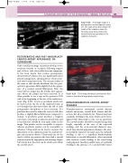
Figure 8.28 A montage image of a postoperative carotid endarterectomy site. The small arrows demonstrate the length of the endarterectomy site.
The large arrow demonstrates the intima-media layer proximal to the endarterectomy site.
Color image showing a carotid artery that has been treated with angioplasty and stenting.
CCA
105
Vein
ICA
POSTOPERATIVE AND POST-ANGIOPLASTY CAROTID ARTERY APPEARANCE ON ULTRASOUND
Only a small percentage of patients develop severe recurrent stenosis or occlusion following surgery and, of these, only a few suffer from any symptoms. It has been shown that routine postoperative ultrasound surveillance does not significantly affect patient management, and patients are often rescan- ned only if symptoms recur. The scan procedure is the same as that already described, but the post- operative appearance differs slightly from the appear- ance of a normal carotid bifurcation. First, the vessel wall no longer has the double layer appear- ance where the plaque has been removed. It is often possible to see a step in the posterior CCA wall at the beginning of the site of the endarterec- tomy (Fig. 8.28). A vein or prosthetic patch may be used to close the site of the endarterectomy, as it is thought that this may reduce the risk of early postoperative thrombosis or late re-stenosis. If a patch has been used to widen the vessel, it will often produce a slightly dilated bifurcation compared to normal. A prosthetic patch produces a brighter echo than a vein patch or adjacent arterial wall, and it can therefore usually be seen quite easily on the image. Vein patches can be susceptible to rupture whereas prosthetic patches can be susceptible to infection. Ultrasound can be used to measure the dimensions of the endarterectomy site and investi- gate any recurrent disease. Ultrasound can also be used to follow up patients who have had a carotid stenosis treated by angioplasty and stenting. Figure 8.29 shows how the stent can be clearly seen within the carotid artery.
Figure 8.29
NONATHEROMATOUS CAROTID ARTERY DISEASES
Nonatheromatous extracranial carotid diseases include aneurysms, carotid body tumors and dis- section, but all are relatively rare. Patients may have a pulsatile swelling in the neck, which can be inves- tigated with ultrasound to rule out an aneurysm. The carotid arteries should be scanned along their length, especially in the area of the suspected swelling, and the cross-sectional diameter mea- sured. Any unusual appearances relating to the arter- ies should be reported. In many cases, the ‘pulsatile swelling’ is due to a superficial brachiocephalic bifurcation or carotid bifurcation, often associated with tortuous vessels, leading to the vessel being easily palpated. Another possible cause of a pulsatile swelling is the presence of a carotid body tumor.
