
Page 116 - Libro vascular I
P. 116
Chap-08.qxd 1~9~04 16:42 Page 107
ULTRASOUND ASSESSMENT OF THE EXTRACRANIAL CEREBRAL CIRCULATION
107
Box 8.2 Information to include in a carotid scan report, especially when surgery is performed on the basis of ultrasound alone
● Locations and appearance of any atheroma seen within the CCA, bifurcation and ICA
● Significant velocity increases seen in the
carotid arteries and an estimation of the
degree of narrowing present
● Abnormal waveforms seen within the CCA,
ICA and ECA
● Endpoint of ICA disease
● Presence and direction of vertebral artery flow
● Level of the carotid bifurcation in relation to
the angle of the jaw
● Limitations of the examination
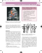
RMCA
LACA LMCA
RPCA P1
P2
LPCA
RACA
Transcranial color flow imaging can be used to investigate the intracerebral circulation (see Fig. 8.2A).
assessment of the cerebral circulation. The tech- niques used in transcranial Doppler assessment are beyond the remit of this book, and the reader is referred to the Further reading section at the end of this chapter.
REPORTING
The ultrasound report should describe the pres- ence, locations and appearance of any atheroma seen within the CCA and ICA. Any significant velocity increases along the carotid arteries should be reported and interpreted to estimate the degree of narrowing present. Abnormal waveforms seen within the CCA, ICA or ECA should also be described, along with a suggestion as to what they may indicate. The presence and direction of verte- bral artery flow should be noted. The report should make it very clear if there was any limitation of the carotid examination, such as the following:
● Inconclusive identification of an occlusion or subocclusion
● Calcification obscuring the vessel for more than 1cm
● No visible endpoint to ICA disease
● Whether the scan was otherwise suboptimal.
Figure 8.32
LR ICA ICA
Example of a diagrammatic method of reporting a carotid ultrasound scan result.
Figure 8.33
When ultrasound is to be used to select patients for surgery, without the use of angiography, it is essential that the examination and report should cover the points listed in Box 8.2. The report can consist of a written report alone or may include images of atheroma and waveforms seen. Alternatively, a diagrammatic representation of the disease seen can be produced. Figure 8.33 is an example of a diagrammatic method of producing a report. It is important that the department has a written protocol, including the criteria used to interpret the Doppler findings and the method of reporting to be used.
