
Page 115 - Libro vascular I
P. 115
Chap-08.qxd 1~9~04 16:42 Page 106
106
PERIPHERAL VASCULAR ULTRASOUND
The carotid body is a small structure within the ves- sel wall, situated at the carotid bifurcation, and is responsible for detecting blood gases and pH. As a carotid body tumor grows, it causes the ICA and ECA to be splayed apart, and small tortuous vessels can often be seen within the tumor with color flow imaging (Fig. 8.30). However, further investigation is required to confirm any ultrasound findings. Carotid artery wall dissection, which can be due to trauma, can create a false lumen within the carotid arteries (Fig. 8.31). This may remain patent and be seen as a second flow lumen on color flow imaging. Alternatively, the false lumen may occlude, causing a reduction in the residual vessel lumen or possibly a complete occlusion of the vessel. An intimal flap may be seen on the image as a fine line within the lumen that may move due to the pulsatile blood flow; however, it may be difficult to image.
TRANSCRANIAL DOPPLER ULTRASOUND
Generally, ultrasound is not easily transmitted through bone, making imaging within the skull dif- ficult. However, the temporal bone is thinner than the rest of the skull and, by using low-frequency ultrasound (e.g., 2 MHz), it is possible to obtain both color flow images and spectral Doppler recordings from segments of some of the intracranial vessels. Nonimaging transcranial Doppler has been used to monitor flow in the MCA during carotid surgery for many years. Carotid endarterectomy involves exposing the carotid bifurcation and clamping the CCA, ICA and ECA. This can lead to compromised cerebral circulation, and, where appropriate, a tem- porary plastic shunt can be used to maintain flow between the CCA and ICA while the plaque is surgically removed. Transcranial Doppler enables MCA blood velocity to be measured, allowing fail- ure of the shunts to be detected. Air and particulate emboli generated at the site of the endarterectomy can be detected, using Doppler ultrasound, as they travel through the MCA. Emboli give a characteris- tic ‘chirp’ on the audible Doppler signal and may appear as a brighter line on the spectral Doppler waveform.
Transcranial color flow imaging is performed using low-frequency phased array transducers and can provide a color map of part or all of the MCA, ACA, PCA and circle of Willis (Fig. 8.32).
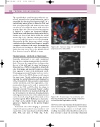
ECA
Transverse image of a carotid body tumor lying between the ICA and ECA.
ICA
Figure 8.30
A
ICA
B
CCA
B-mode image of a carotid artery wall dissection showing a false lumen imaged in transverse
section (A) and in longitudinal section (B).
Figure 8.31
However, views can sometimes be limited by atten- uation caused by the temporal bone. Transcranial color flow imaging requires an in-depth under- standing of possible collateral pathways and, as yet, does not have a clear role in routine ultrasound
