
Page 128 - Libro vascular I
P. 128
Chap-09.qxd 29~8~04 14:46 Page 119
at short notice. Bowel preparations have proved use- ful, although in practice they can be difficult to administer to elderly or diabetic patients and are impractical in a single visit clinic.
The patient should have an empty bladder prior to an aortoiliac scan as this improves the visualization of these segments and also causes less patient dis- comfort if transducer pressure has to be applied. The examination room should be at a comfortable ambient temperature (20°C) to avoid peripheral vasoconstriction.
Scanner setup
A peripheral arterial scanning option should be selected before starting the examination, but adjustment of the control settings will often be required in the presence of significant disease (see Ch. 7). The color PRF is usually set in the 2.5–3 kHz range for demonstrating moderately high velocity flow.
STARTING THE SCAN
It is useful to start the assessment by examining the CFA at the groin, as the observed blood flow patterns at this level can reveal information about the condition of the aortoiliac arteries and also pro- vide some clues to the condition of the superficial femoral artery (SFA) (i.e., origin occlusion or high resistance flow pattern due to proximal obstruction). It is important to have a good understanding of the anatomy of the arteries and veins at the level of the groin and to be able to identify the major branches and junctions and their relationship to each other (Fig. 9.6). A 5 MHz, or broad-band equivalent, linear array transducer is the most suitable probe for scanning the femoral, popliteal and calf arteries. A 3.5 MHz, or broad-band equivalent, curved linear array abdominal transducer is used for the aortoiliac segment. The segmental guidelines can be used in any order. A combination of B-mode imaging, color flow imaging and spectral Doppler recordings should be used throughout the examination. Color flow imaging is essential for identifying the aortoiliac and calf arteries. Spectral Doppler velocity measure- ments should be made at an angle of 60° or less (see p. 69).
DUPLEX ASSESSMENT OF LOWER LIMB ARTERIAL DISEASE
119
Assessment of the aortoiliac artery
and CFA
The patient should be relaxed and lying in a supine position with the head supported by a pillow. The patient should be asked to relax the abdominal mus- cles and to rest the arms by the sides. The scanning positions for assessing the inflow arteries are shown in Figure 9.7, and a color image of the arteries is shown in Figure 9.8. The procedure for assessment is as follows:
1. Usinga5MHz,orbroad-bandequivalent,linear array transducer, the CFA is identified at the level of the groin in transverse section, where it lies lat- eral to the common femoral vein (Figs 9.6 and 9.7A). The CFA is then followed proximally in longitudinal section until it runs deep under the inguinal ligament and can no longer be assessed
CIA IIA EIA
CFA
Aorta
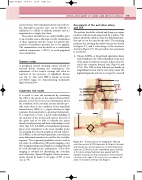
Figure 9.7
C
B
Aorta CIA CIA
EIA
IIA
A CFA Vein
D SFA
CFA
CIA
EIA IIA
Probe positions for imaging the CFA and aortoiliac arteries. A: CFA transverse. B: Origin of external and internal iliac arteries transverse. C: Aortic bifurcation transverse. D: Arteries in the longitudinal plane. Starting at the groin and pushing bowel gas upward with the transducer (arrow) can help visualization. Positioning the color box to the edge of the scan sector can improve the angle of insonation with spectral Doppler.
