
Page 130 - Libro vascular I
P. 130
Chap-09.qxd 29~8~04 14:46 Page 121
PA
POP
TPT
AT
SFA
DUPLEX ASSESSMENT OF LOWER LIMB ARTERIAL DISEASE
121
PA
PT
PER
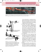
A color montage of the femoropopliteal and calf arteries. The image shows the profunda femoris artery (PA), SFA, popliteal artery (POP), tibial peroneal trunk (TPT), PT, AT and peroneal artery (PER).
Figure 9.9
often be followed for a considerable distance, particularly if the SFA is occluded and it is sup- plying a collateral pathway to the lower thigh. The origin of the SFA is usually located antero- medial to the profunda femoris artery, requiring a slight inward turn of the transducer.
3. The SFA is then followed distally along the medial aspect of the thigh in a longitudinal plane, where it will lie above the superficial femoral vein (Fig. 9.10C). If the image of the SFA is lost it is easier to relocate in transverse section (Fig. 9.10D). In its distal segment the SFA runs deep and enters the adductor canal, becoming the popliteal artery. It is usually possible to image the proximal popliteal artery to just above the knee level from this position (see Fig. 9.10E). A 3.5 MHz transducer can help to image the artery in a large thigh.
4. The popliteal artery can be examined by rolling the patient onto the side. Alternatively, the patient can lie in a prone position, resting the foot on a pillow, although a lot of elderly patients are not able to tolerate this position. It is also possible to image the popliteal artery with the legs hanging over the edge of the examination table and the feet resting on a stool. Whichever method is used, it is important not to overex- tend the knee joint as this can make imaging difficult.
5. Starting in the middle of the popliteal fossa, the popliteal artery is located in transverse sec- tion and is seen posterior to the popliteal vein (Fig. 9.10F). Turning into a longitudinal plane, the popliteal artery is then followed proximally,
Artery
Vein
Vein Artery
B
CFA
A
PA
SFA D
POP F
SFA
Profunda
Femoral artery vein
Transducer in transverse position behind knee in popliteal fossa
E
AT
TPT
C
Medial aspect of thigh
Figure 9.10 Probe positions for imaging the femoropopliteal arteries. A: Femoral artery bifurcation transverse. B: Femoral bifurcation longitudinal. C: SFA longitudinal. D: SFA transverse. E: Proximal popliteal artery above-knee longitudinal. F: Popliteal artery transverse. G: Popliteal artery longitudinal, from the popliteal fossa. H: Origin of the AT.
CFA
SFA
POP
H G
