
Page 131 - Libro vascular I
P. 131
Chap-09.qxd 29~8~04 14:46 Page 122
122
PERIPHERAL VASCULAR ULTRASOUND
above the popliteal fossa, to overlap the area previously examined from the lower medial thigh (Fig. 9.10G).
6. The popliteal artery is then examined longitudi- nally across and below the popliteal fossa, where it is possible to continue directly into the tibioper- oneal trunk. The tibioperoneal trunk can be imaged from a number of positions.
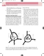
Assessment of the tibial arteries
The tibial arteries can be imaged from several differ- ent transducer positions, as demonstrated in Figure 9.11. It is often easier to locate the tibial arteries in the distal calf and follow them proximally to the top of the calf. However, for the purposes of this section, the description of the examination starts just below the knee. It should be noted that imag- ing of the distal tibial arteries at the ankle is often easier with a high-frequency 10MHz, or broad- band equivalent, flat linear array transducer.
Anterior tibial artery
1. Withthelegrolledoutwardandthekneeslightly flexed, the origin of the anterior tibial (AT) artery is imaged from a posteromedial position just below the knee, where it will be seen to drop immediately away from the popliteal artery (Fig. 9.10H). Often it is only possible to see the first 1–2cm of the AT from this position. The tibioperoneal trunk is usually seen as a direct continuation of the popliteal artery distal to the AT artery origin.
2. The proximal AT artery is then imaged from the anterolateral aspect of the upper calf, just below the knee, where it will be seen to rise toward the transducer in a curve, through the interosseous membrane. The membrane can be identified as a bright echogenic line running between the tibia and fibula in cross section. The artery will lie on top of the membrane. The AT artery is then followed distally, along the anterolateral
AT
LM
AT PER
AE TATDL M
ETI RA AL LF
TPT
TPT
TPT AB
AE TD E AT I RTA AL
F
PER PT
L TPT
PT PER
Figure 9.11 Cross-sections of the calf to show longitudinal transducer positions for imaging the tibial arteries and veins in the calf. A: Several positions can be used to image the vessels in the upper calf proximal to the bifurcation of the tibioperoneal trunk (TPT). B: Probe positions to image the PT, AT and peroneal artery (PER) in the mid- and lower calf. Note that it is possible to image two vessels from a similar position, as shown.
PER
