
Page 133 - Libro vascular I
P. 133
Chap-09.qxd 29~8~04 14:46 Page 124
124
PERIPHERAL VASCULAR ULTRASOUND
Table 9.4 Common problems encountered during duplex evaluation of the lower limb arteries
Segment
Aortoiliac arteries
Aortoiliac arteries
Femoropopliteal arteries Femoropopliteal arteries
Tibial arteries
Tibial arteries
Problem
Bowel gas obscuring part or all of the image
Tortuous arteries
Severe calcification of the artery producing color image dropout
Obese patient with large thigh
Large calf with gross edema
Very low flow due to proximal occlusions
Solutions
Try different probe positions (medial, lateral or coronal positions); leave the segment and try again in a few minutes
Use the color display to follow the artery; considerable adjustment of the probe position is often needed
Try different transducer positions to work around the calcification
When using a broad-band transducer, lower the color and spectral Doppler transmit frequencies for better penetration; consider switching to a 3.5 MHz curved linear array transducer in very difficult situations
Start the scan at the ankle and work proximally; a 3.5 MHz linear array probe can be used to image these vessels proximally
Lower the pulse repetition frequency and wall filters; place the leg in a dependent position to increase distal blood flow
SCAN APPEARANCES
B-mode images
Normal appearance
Like the carotid arteries, the lumen of a normal peripheral artery should appear clear, and the walls should be uniform along each arterial segment, although noise may cause speckle within the image of the vessel. The intima-media layer of the arterial wall is sometimes seen in normal femoral and popliteal arteries. In practice, it is frequently difficult to clearly image the vessels in the aortoiliac segment, abductor canal region and calf without the help of color flow imaging.
Abnormal appearance
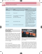
Areas of atheroma, particularly if they are calcified, may be seen within the vessel lumen. The atheroma may be extensive and diffusely distributed, especially in the SFA (Fig. 9.12). Large plaques at the common femoral bifurcation are relatively easy to image, and
CALCIFICATION
Figure 9.12 Calcified atheroma (arrows) is present in the SFA, leading to drop-out of the color flow signal in parts of the lumen.
these may extend into the proximal profunda artery or SFA. Calcification of the arterial wall, especially in diabetic patients, produces strong ultrasound reflections, and the walls of the calf arteries can appear particularly prominent (Fig. 9.4). When an arterial segment has been occluded for some time, the vessel may contract and appear as a small cord adjacent to the corresponding vein. This appearance
