
Page 135 - Libro vascular I
P. 135
Chap-09.qxd 29~8~04 14:46 Page 126
126
PERIPHERAL VASCULAR ULTRASOUND
CIA CIV
EIA
PA
AB
IIA
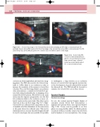
Figure 9.15 A: Color flow image of the femoral bifurcation demonstrating an SFA origin occlusion (arrow). The
profunda femoris artery (PA) is patent. B: Color flow image of an external iliac artery (EIA) occlusion (arrow). The CIA and internal iliac artery (IIA) are patent. The common iliac vein (CIV) is visible in this image.
occlusion in both longitudinal and transverse imag- ing planes. The PRF often needs to be lowered (typ- ically to 1kHz) distal to an occlusion in order to increase the sensitivity of the scanner to lower flow velocities. The color flow image distal to an occlusion often demonstrates a continuous forward flow pat- tern with reduced pulsatility due to damping of the normal blood flow pattern. Blood flow in the main artery may also improve progressively over the first few centimeters distal to the occlusion as more col- lateral vessels join the main trunk. This effect can be observed on the color flow image (Fig. 9.17). High- velocity flow in a collateral vessel can produce an area of marked color flow disturbance in the main artery at the point where the collateral joins. This can be misinterpreted as a stenosis. Spectral Doppler should be used to interrogate this area carefully. It is possible
Figure 9.16 A short mid-SFA occlusion is demonstrated by an absence of color flow in the vessel (large arrow). Large collateral vessels are seen at both ends of the occlusion (small arrows).
to misdiagnose a long stricture as an occlusion because of very slow flow through the stricture due to the development of good collateral flow around the diseased site. The PRF should be lowered to examine low-velocity flow across these lesions.
Spectral Doppler
Normal recordings
At rest, the normal spectral Doppler display of extremity arterial blood flow demonstrates a triphasic flow pattern with a clear spectral window (Fig. 9.18). It may even be possible to see four phases in young healthy adults. In elderly patients or patients with poor cardiac output, the waveform may be biphasic or even monophonic. The average
