
Page 134 - Libro vascular I
P. 134
Chap-09.qxd 29~8~04 14:46 Page 125
DUPLEX ASSESSMENT OF LOWER LIMB ARTERIAL DISEASE
125
POP A
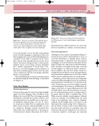
Figure 9.13 An acute occlusion of the popliteal artery. The vessel is patent to the level of the two arrows. The occlusion is demonstrated by the relatively low level echoes in the lumen distally. Note some intimal detail
is still visible in the occluded section (curved arrow).
Figure 9.14 Two severe stenoses are demonstrated in the SFA by areas of color flow disturbance and aliasing (arrows).
demonstrated as continuous flow in one color scale but there should be no evidence of arterial stenosis.
Abnormal appearance
Utilizing the color controls as described in Chapter 7, arterial stenoses will be demonstrated as areas of color flow disturbance or aliasing. Severe stenoses frequently produce a disturbed color flow pattern extending 3 to 4 vessel diameters beyond the lesion (Figs 9.8 and 9.14). Any areas of color flow distur- bance should be investigated with angle-corrected spectral Doppler to estimate the degree of narrow- ing. In addition, the color flow image of flow in a nondiseased artery distal to severe proximal disease may demonstrate damped low-velocity flow, which will be seen as continuous flow in one direction.
Occlusions of lower limb arteries most frequently occur in the SFA and popliteal artery. An occlusion is demonstrated by a total absence of color flow in the vessel. Occlusions can occur at the origins of arteries or in mid-segment. If an artery is occluded from its origin, at the level of a major bifurcation, flow will normally still be seen in the sister branch. For exam- ple, the profunda femoris artery is usually found to be patent when the SFA is occluded (Fig. 9.15). When an artery occludes in mid-segment, collateral vessels are normally seen dividing from the main trunk at the beginning of the occlusion. Similarly, collateral vessels resupply flow to the artery at the distal end of the occlusion (Fig. 9.16). Collateral ves- sels can follow tortuous routes as they divide from the main trunk, and they are sometimes only seen when the main artery is imaged in cross-section. It is therefore helpful to interrogate any suspected
is most frequently seen in the SFA and popliteal artery. B-mode imaging in combination with color flow imaging is also very useful for identifying acute occlusions of the SFA or popliteal artery, where there may be fresh thrombus present in the vessel lumen. The lumen will appear clear or demonstrate minimal echoes on the image, because thrombus has a similar echogenicity to blood (Fig. 9.13). However, color flow imaging reveals an absence of flow in the occluded segment of the vessel. The start of the occlusion can often be very abrupt, with little disease seen proximally.
Abnormal dilatations or arterial aneurysms should be measured using the B-mode image, as described in Chapter 11.
Color flow images
Normal appearance
Normal arterial segments can be interrogated rapidly using color flow imaging. There should be color filling to the vessel walls. The color image normally demonstrates a pulsatile flow pattern, with the color alternating between red and blue due to flow rever- sal during the diastolic phase (see Ch. 5). There are situations in which flow in nondiseased lower limb arteries may have reduced pulsatility or even be continuous. Examples include increased flow (hyperemia) due to limb infection or the presence of arteriovenous fistulas. Hyperemic flow will be
