
Page 139 - Libro vascular I
P. 139
Chap-09.qxd 29~8~04 14:46 Page 130
130
PERIPHERAL VASCULAR ULTRASOUND
syndrome, the patient should lie prone with the legs gently flexed and the feet hanging over the end of the examination table. The below-knee popliteal artery should be imaged at the level of the gastroc- nemius muscle heads. The patient should point the foot down (plantar flex) against a counterpressure, typically by having a colleague apply moderate pres- sure against the foot. Narrowing or occlusion of the popliteal artery during this maneuver may indicate popliteal entrapment syndrome. However, there is evidence to suggest that significant compression of the popliteal artery can occur in normal volunteers during this investigation, casting some doubt on the usefulness of this test (Erdoes et al 1994).
Cystic adventitial disease of the
popliteal artery
This rare disease is caused by cystic swelling of the arterial wall, which impinges into the lumen of the popliteal artery, leading to eventual occlusion. The location of the lesion is often found across the knee joint. It should be considered as a potential cause of symptoms in the young patient, especially in the absence of any other pathology. Treatment is by excision and local repair or bypassing.
REPORTING
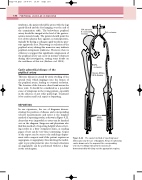
In our experience, the use of diagrams demon- strating the position of disease and corresponding velocity measurements and ratios is the simplest method of reporting results, as shown in Figure 9.22. Areas that were impossible to assess can be hatched out on the diagram. Surgeons and physicians also find this method of reporting helpful when review- ing results in a busy outpatient clinic, as reading pages of text can be very time-consuming. Copies of the report can be sent to the radiology depart- ment with a request card if the patient requires an angiogram or angioplasty, thus allowing the radiol- ogist to pre-plan puncture sites. In many situations an angioplasty can be performed without a diag- nostic arteriogram.
CIA stenosis 8 × velocity increase
CIA occlusion
SFA occlusion
Severe diffuse SFA disease
Tibial artery disease
The easiest method of reporting lower limb scans is by the use of diagrams. Areas of narrowing
Figure 9.22
can be drawn onto the map and the corresponding velocity recordings indicated. Occlusions are demonstrated by blocking out the appropriate regions.
