
Page 145 - Libro vascular I
P. 145
Chap-10.qxd 29~8~04 14:48 Page 136
136
PERIPHERAL VASCULAR ULTRASOUND
arteries. In addition, the thoracic outlet can be investigated for possible compression of the SA. A minimum of half an hour should be allocated for the examination.
There is no special preparation required prior to the scan, although the patient will have to expose the shoulder and upper arm for scanning of the distal SA and axillary arteries. The examination room should be at a comfortable ambient temperature (20° C) to prevent vasoconstriction of the distal arteries. The patient should lie supine with the head supported on a thin pillow for comfort. The SA and proximal axillary artery can be scanned by sitting behind the patient. This is usually a more comfortable position than scanning from the side of the patient. To image the distal axillary and brachial arteries, the patient should be examined from the side of the examination table and the arm should be abducted, be externally rotated and be resting on an arm board or a suitable rest. The dis- tal brachial, radial and ulnar arteries are imaged with the hand in a palm-up position, resting on a support. The scanner should be configured for a peripheral arterial examination, and in the absence of a specific upper limb preset, a lower limb arterial option should be selected.
SCANNING TECHNIQUES
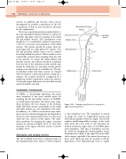
A 5MHz, or broad-band equivalent, flat linear array transducer is the most suitable probe for scanning the SA and axillary arteries. A 10MHz, or broad-band equivalent, flat linear array trans- ducer produces the best images of the brachial, radial and ulnar arteries, particularly as the radial and ulnar arteries are very superficial at the wrist. In addition, a 5–7MHz curved linear array trans- ducer can be useful for imaging the proximal SA at the level of the supraclavicular fossa, as it fits more easily into the contour of this region. The trans- ducer positions for imaging the upper extremity arteries are shown in Figure 10.3. A color flow montage of the upper extremity arteries is shown in Figure 10.4.
Subclavian and axillary arteries
The SA is initially located in a transverse plane in the supraclavicular fossa, where it will lie superior
Supraclavicular fossa Clavicle
Infraclavicular fossa Axillary artery
Brachial artery
Radial artery
Subclavian artery
Transducer positions for scanning the upper extremity arteries.
Figure 10.3
to the subclavian vein. The transducer is turned to image the artery in longitudinal section and followed proximally toward its origin. The left SA origin is usually impossible to image, as the vessel arises from the aortic arch. It can sometimes be tracked toward its origin with a 2–2.5 MHz phase array transducer. This type of transducer can also be useful for imaging the brachiocephalic artery. Sometimes the origin of the right SA can be diffi- cult to image, especially if the patient has a large or short neck. Extra gel may be needed to fill the depression of the supraclavicular fossa to enable
Ulnar artery
