
Page 146 - Libro vascular I
P. 146
Chap-10.qxd 29~8~04 14:48 Page 137
DUPLEX ASSESSMENT OF UPPER EXTREMITY ARTERIAL DISEASE
137
below the clavicle (see Fig. 10.12). A mirroring artifact of the SA is often seen due to the chest wall beneath the artery (see Fig. 7.7).
The SA reappears from underneath the clavicle and is followed distally, where it becomes the axillary artery. Two positions may be used to image the length of the axillary artery. The first is the anterior approach, in which the axillary artery will be seen to run deep beneath the shoulder muscles. A 3.5 MHz curved array transducer can sometimes be useful for following the distal axillary artery from this position. The second approach images the axillary artery from the axilla (armpit), where it can be followed distally to the brachial artery.
It is worth noting that the proximal segment of the internal thoracic artery, a proximal branch of the SA, can often be imaged. This artery is fre- quently used in coronary bypass surgery and is sur- gically grafted to the heart. It divides at a 90° angle from the inferior aspect of the SA to run down the chest wall. It is possible to confirm graft patency by identifying flow in the proximal thoracic artery just beyond its origin. The flow pattern in the artery supplying the heart will exhibit an unusual wave- form shape, as most of the flow occurs in the dias- tolic phase of the cardiac cycle.
Brachial artery
The brachial artery is followed as a continuation of the axillary artery along the inner aspect of the arm to the elbow, where it curves around to the cubital fossa and lies in a superficial position.
The distal brachial artery is scanned across the elbow to the point where it divides in the upper forearm into the radial and ulnar arteries.
Radial and ulnar arteries
The bifurcation of the brachial artery into the radial and ulnar arteries is easier to locate in a transverse plane. The two arteries are then followed distally to the wrist in a longitudinal plane. In its proximal segment, the ulnar artery runs deep to the radial artery before becoming more superficial in the mid- forearm. It is often easier to locate the radial and ulnar arteries at the wrist and then to follow them back to the elbow.
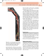
SA
AA
DB
BA
A color flow montage of the left upper extremity arteries demonstrating the subclavian artery
Figure 10.4
UA
RA
I
(SA), axillary artery (AA), brachial artery (BA), deep brachial artery (DB), radial artery (RA), common interosseous artery (I) and ulnar artery (UA).
good contact with a flat linear array transducer. The SA should then be followed laterally in longi- tudinal section, where it will disappear underneath the clavicle. There will be a large acoustic shadow
