
Page 147 - Libro vascular I
P. 147
Chap-10.qxd 29~8~04 14:48 Page 138
138
PERIPHERAL VASCULAR ULTRASOUND
Palmar arch and digital arteries
Duplex scanning can be used to image the palmar arch and digital vessels, although continuous wave Doppler can be considerably quicker and easier to use for the detection of arterial signals, especially in the digital arteries. The radial artery is sometimes used as a graft for coronary artery bypass surgery. It is possible to listen to the digital arteries and palmar arch flow signals with continuous wave Doppler, while the radial artery is being manually compressed, to ensure that perfusion to the hand and fingers is being maintained by the ulnar artery. If this is not the case, removal of the radial artery could result in hand ischemia.
Commonly encountered problems
Most problems occur due to poor imaging, especially in large or obese patients, in whom the proximal arteries may be very difficult to image. In particular, the SA in the area of the supraclavicular fossa can be difficult to locate. Color flow imaging can present a confusing display as there are often strong signals from the adjacent subclavian vein, which may appear pulsatile due to the proximity to the right side of the heart. Imaging of the axillary artery can be dif- ficult where the artery runs deep under the shoul- der muscles. Scanning from the axilla or selecting a lower frequency probe may help.
ULTRASOUND APPEARANCE
Normal appearance
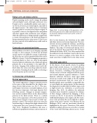
The normal appearance of upper extremity arteries is the same as that described for the duplex scan- ning of lower limb arteries (see Ch. 9). The spec- tral Doppler waveform is normally triphasic at rest but becomes hyperemic with high diastolic flow following exercise. Changes in external tempera- ture can have marked effects on the observed flow patterns in the distal arteries. There is a cyclical effect on the appearance of the flow patterns in the distal arteries related to factors such as body tem- perature control. This cyclical effect can cause the waveform shape to change from high-resistance flow to hyperemic flow over a period of a minute or two (Fig. 10.5). Peripheral vasodilation will cause a reduction in peripheral resistance and an increase in
A cyclical change in the appearance of the blood flow patterns in the radial and ulnar arteries can
be observed, relating to factors such as the control of body temperature.
Figure 10.5
flow. In this situation, the waveform in the radial and ulnar arteries can become hyperemic. Vasocon- striction increases peripheral resistance, producing a reduction in flow, and the waveform becomes biphasic. The range of normal peak systolic veloci- ties in the SA has been reported as 80–120cm/s (Edwards & Zierler 1992). It is often assumed that the radial artery is the dominant vessel in the forearm because it is easier to palpate at the wrist, but in many cases there is higher flow in the ulnar artery.
Abnormal appearance
In the absence of any specific criteria for grading upper limb arterial stenoses, we would advocate the use of the same criteria as for grading lower limb disease. Therefore, a doubling of the peak systolic velocity across a stenosis compared with the proxi- mal normal adjacent segment indicates a 50% diameter reduction. However, many upper limb lesions are located at the origin to the SA, making proximal measurements from the aortic arch or brachiocephalic artery unreliable or impossible due to vessel depth, size and geometry. In this situation the diagnosis is usually made by indirect signs, such as high-velocity jets, turbulence or post-stenotic damping (Fig. 10.6). In addition, the ipsilateral vertebral artery should be examined for evidence of flow changes, indicated by damping or flow reversal (see Ch. 8). It can also be very difficult to visibly identify plaques at the origin to the SA. Occlu- sions of the proximal SA can be difficult to differ- entiate from severe stenoses (von Reutern & von Büdingen 1993), and any uncertainty should be highlighted in the report. Dissection of the radial, brachial or axillary arteries can occur due to trauma of the vessel wall following catheter access. It may
