
Page 148 - Libro vascular I
P. 148
Chap-10.qxd 29~8~04 14:48 Page 139
DUPLEX ASSESSMENT OF UPPER EXTREMITY ARTERIAL DISEASE
139
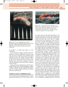
Figure 10.7 An embolus from the heart has acutely obstructed the brachial artery at the elbow. The arterial lumen appears clear, as the embolus has a similar echogenicity to blood, but there is a sudden cessation of flow at the start of the occlusion (arrow).
the region where the SA and brachial plexus leave the chest and pass in between the anterior and middle scalene muscles over the first rib and under- neath the clavicle (Fig. 10.8). This is a compact anatomical area, and compression on the nerves or arteries by a number of mechanisms can produce sensory symptoms in both the hand and arm. Compression can occur in three main areas. The first is at the point where the SA passes between the sca- lene muscles and can be caused by muscle hypertro- phy or fibrous bands or may be due to the presence of an additional accessory rib originating from the seventh thoracic vertebra, termed a cervical rib (Fig. 10.9). Accessory ribs occur in less than 1% of the population (Makhoul & Machleder 1992). The second area of compression occurs as the artery runs between the first rib and clavicle. Fibrous bands or fibrosis due to injuries in this region, such as fractures of the clavicle, can also cause compres- sion. The third, less common area of compression occurs in the subcoracoid region, where the axil- lary artery runs under the pectoralis minor muscle and close to the coracoid process of the scapula.
Typically, the vessels and nerves are compressed when the arm is placed in specific positions. The symptoms include sensory changes, such as pain, pins and needles in the hand, hand weakness and other neurological disorders. TOS can be purely neurogenic, due to compression of the brachial plexus alone (this accounts for approximately 90% of cases). Neurogenic TOS often produces abnormal nerve conduction recordings and can be associated
Figure 10.6
A severe high-grade stenosis of the proximal SA (arrow) is demonstrated by marked color flow disturbance and aliasing, high peak systolic velocity
(389 cm/s), abnormal waveform shape and spectral broadening.
be possible to see flaps, dual lumens or acute obstruction.
Acute occlusions of upper extremity arteries are frequently caused by embolization from the heart and occur most commonly in the brachial, radial and ulnar arteries. The arterial lumen may appear relatively clear, but there will be an absence of flow in the vessel as demonstrated by color flow imag- ing (Fig. 10.7). Some acute occlusions occur as a result of embolization from the SA due to damage caused by TOS.
Large arteriovenous malformations will be imme- diately obvious with color flow imaging as a region of high vascularity. Spectral Doppler will demon- strate low-resistance, high-volume flow waveforms within the malformation.
THORACIC OUTLET SYNDROME (TOS)
The vascular laboratory is frequently asked to assess patients with suspected TOS. The thoracic outlet is
