
Page 149 - Libro vascular I
P. 149
Chap-10.qxd 29~8~04 14:48 Page 140
140
PERIPHERAL VASCULAR ULTRASOUND
These aneurysms can be the source of distal emboli in the fingers, which can be the initial presentation of a patient with TOS. There is still considerable debate about the assessment and treatment of TOS, which often involves surgical resection of a cervical rib and sometimes the first rib, with the division of any fibrous bands to relieve the com- pression. Although the majority of patients who have undergone surgery show improvement in symptoms, a few show no signs of improvement and may return to the vascular laboratory for fur- ther assessment.
Maneuvers for assessing TOS
Continuous wave Doppler recording of the radial artery signal, performed with the arm in a range of positions, can be a useful prelude to the duplex examination (Fig. 10.10). There are a range of provocation maneuvers that can be used, but the most common include the following.
Hyperabduction test
The patient should be sitting comfortably, and the arm should then be slowly extended outward (abducted). With the arm fully abducted, the fore- arm is rotated so that the palm faces upward and the elbow downward (external rotation). The arm should be raised and lowered in this position and the patient’s head turned away from the side under investigation. This test can indicate compression between the clavicle and first rib or coracoid region.
Costoclavicular maneuver
The patient is asked to push the chest outward while forcing the shoulders backward with deep inhalation, the so-called ‘military position’, as this may reveal arterial compression between the clavi- cle and first rib.
Deep inspiration maneuver
During deep inspiration the patient is asked to extend the neck and rotate the head to the affected side and then to the other side while the pulse is checked at the wrist. A positive test indicates
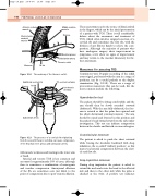
Anterior scalene muscle
Sternocleido- mastoid muscle
Sternum
Figure 10.8
Middle scalene muscle
Clavicle
Brachial plexus Subclavian artery
Thoracic outlet
Additional cervical rib Clavicle 1st rib
Subclavian artery
7th cervical vertebra
1st thoracic vertebra
1st rib
The anatomy of the thoracic outlet.
Sternum
Figure 10.9 The presence of a cervical rib originating from the seventh thoracic vertebra can cause compression of the brachial nerve plexus and subclavian artery.
with muscle weakness and wasting in the lower arm or hand.
Arterial and venous TOS is less common and accounts for approximately 10% of cases, although there is sometimes a combination of neurogenic and vascular compression. Aneurysmal dilations of the SA are sometimes seen just distal to the point of compression due to post-stenotic dilation.
