
Page 16 - Libro vascular I
P. 16
ULTRASOUND AND IMAGING 7
will be seen later), but ultrasound systems usually make an estimate by assuming that the speed of sound is the same in all tissues: 1540 m/s. This can lead to small errors in the estimated distance travelled because of the variations in the speed of sound in different tissues.
GENERATION OF ULTRASOUND WAVES
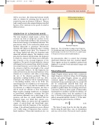
The term transducer simply means a device that converts one form of energy into another. In the case of an ultrasound transducer, this conversion is from electrical energy to mechanical vibration. The piezoelectric effect is the method by which most medical ultrasound is generated. Piezoelectric materials will vibrate mechanically when a varying voltage is applied across them. The frequency of the voltage applied will affect the frequency with which the material vibrates. The thickness of the piezoelectric element will determine the frequency at which the element will vibrate most efficiently; this is known as the resonant frequency of the transducer. The speed of sound within the element will depend on the material from which it is made. A resonant frequency occurs when the thickness of the element is half the wavelength of the sound wave generated within it. At this frequency, the reflected waves from the front and back faces of the element act to reinforce each other, so increasing the size of the vibration produced. When an appropriate coupling medium is used (e.g., ultra- sound gel), this vibration will be transmitted into a surrounding medium, such as the body. The named frequency of a transducer is its resonant fre- quency. This is not to say that the transducer will not function at a different frequency, but that it will be much less efficient at those frequencies. Many modern imaging transducers are designed as broad-band transducers, meaning that they will function efficiently over a wide range of fre- quencies, and these are usually labelled with the frequency range over which they operate (e.g., 4–7MHz). Figure 2.2 shows how the transducer output of narrow-band and broad-band trans- ducers varies with the frequency of the excitation voltage. A broad-band transducer is more efficient over a wider range of frequencies than a narrow- band transducer. Ultrasound transducers also use
Broad-band transducer Narrow-band transducer
Maximum
Frequency
Resonant frequency
Plot of transducer output versus frequency for a broad-band and a narrow-band transducer. A broad- band transducer will be more efficient over a wider range of frequencies than a narrow-band transducer.
the piezoelectric effect to convert the returning ultrasound vibrations back into electrical signals. These signals can then be amplified, analyzed and displayed to provide both anatomical images and flow information.
Pulsed ultrasound
Simple Doppler systems operate with a continuous single-frequency excitation voltage, but all imaging systems and pulsed Doppler systems use pulsed excitation signals. If ultrasound is continuously transmitted along a particular path, the energy will also be continuously reflected back from any boundary in the path of the beam, and it will not be possible to predict where the returning echoes have come from. However, when a pulse of ultrasound is transmitted it is possible to predict the distance (d) of a reflecting surface from the transducer if the time (t) between transmission and reception of the pulse is measured and the velocity (c) of the ultra- sound along the path is known, as follows:
tc
d d 2 2 ( 2 . 3 )
The factor 2 arises from the fact that the pulse travels along the path twice, once on transmission and once on its return. This can be used to predict
Figure 2.2
Chap-02.qxd 29~8~04 13:19 Page 7
Transducer output
