
Page 17 - Libro vascular I
P. 17
Chap-02.qxd 29~8~04 13:19 Page 8
8
PERIPHERAL VASCULAR ULTRASOUND
Time
A B
Time
C D
Time
E F
Time
G H
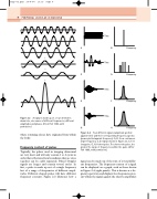
A signal is made up of, or can be broken down into, sine waves of different frequencies, different amplitudes and phases. (From Fish 1990, with permission.)
where returning echoes have originated from within the body.
Frequency content of pulses
Typically, the pulses used in imaging ultrasound are very short and will only contain 1 to 3 cycles in order that reflections from boundaries that are close together can be easily separated. Pulsed Doppler signals are longer and contain several cycles. In fact, a pulse is made up not of a single frequency but of a range of frequencies of different ampli- tudes. Different shaped pulses will have different frequency contents. Figure 2.3 illustrates how a
Frequency
Frequency
Frequency
Frequency
Four different signals (amplitude plotted against time) and their corresponding frequency spectra (power plotted against frequency). A, B: For a continuous single frequency. C, D: Signal shown in Figure 2.3. E, F: A long pulse. G, H: A short pulse. The shorter the pulse, the greater the range of frequencies within the pulse. (After Fish 1990, with permission.)
Figure 2.3
Figure 2.4
signal can be made up of the sum of several differ- ent frequencies. The frequency content of a signal can be displayed on a graph, such as those shown in Figure 2.4 (right panels). This is known as a fre- quency spectrum and displays the frequencies pres- ent within the signal against the relative amplitudes
