
Page 178 - Libro vascular I
P. 178
Chap-12.qxd 29~8~04 14:52 Page 169
ANATOMY OF THE LOWER LIMB VENOUS SYSTEM AND ASSESSMENT OF VENOUS INSUFFICIENCY
169
referred to as the ‘gatekeeper’, as it prevents venous reflux into the proximal calf. The deep veins in the calf contain numerous valves. The LSV and SSV contain approximately 8–10 valves along their main trunks (Browse et al 1999). In addition, there are normally valves at the junctions between the super- ficial and deep veins, and valves protecting many perforating veins so that flow is directed from the superficial venous system to the deep system. In very rare cases, patients may have absent venous valves due to congenital valve aplasia (Eifert et al 2000). This can be the cause of deep venous reflux in the very young patient.
Flow patterns in the venous system
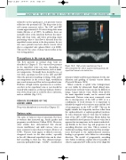
The flow patterns in normal deep veins are described in Chapter 5. The venous flow patterns in the superficial veins can vary, depending on patient position and external factors, such as ambi- ent temperature. Normally there should be no, or very little, spontaneous flow in the LSV and SSV when the patient is standing or sitting. If the ambi- ent temperature in the room is high, vasodilation may result in increased flow in the superficial veins. Evidence of high-volume spontaneous or continu- ous flow in the superficial veins at rest should be treated with suspicion, as this may indicate obstruc- tion of the deep veins or could be due to infection, such as cellulitis (Fig. 12.9).
VENOUS DISORDERS OF THE
LOWER LIMBS
Deep vein thrombosis is covered in Chapter 13.
Varicose veins
The cause of varicose veins is uncertain, but there is evidence that increased age, female gender and pregnancy are risk factors (Callam 1994). However, other studies have demonstrated that chronic venous insufficiency and mild varicose veins are more common in men than in women (Evans et al 1999). Varicose veins appear as dilated, tortuous, elongated vessels on the skin surface, especially in the calf. Abnormal superficial veins can be classi- fied according to their size. The clinical-etiologic- anatomic-pathophysiologic (CEAP) classification
High-volume spontaneous flow is demonstrated in the LSV of a patient with popliteal and
superficial femoral vein obstruction.
Figure 12.9
system is widely used by many clinicians for the clas- sification and grading of chronic venous disease (Porter & Moneta 1995).
Minor cosmetic or spider veins (telangiectases) are not visible by ultrasound. Small dilated intra- dermal veins (reticular veins) can also be difficult to image. Larger varicose veins, which often involve the main superficial trunks, can be assessed with ultrasound. Varicose veins most commonly occur due to incompetence of the LSV or SSV, or to a combination of both systems. It is important to identify the supply to the varicose areas and the level of incompetence in the LSV or SSV. This can be highly variable but frequently involves reflux from the saphenofemoral or saphenopopliteal junctions. In some situations varicose veins may be indepen- dent of the LSV or SSV systems. Much debate has surrounded the development of varicose veins, but it appears that incompetence in the main trunks devel- ops distally, in the lower leg, and progresses in an ascending direction over time. This is contrary to traditional teaching, which proposed that incompe- tence developed at the saphenofemoral junction, leading to progressive valve failure in a descending direction. The model of progressively ascending valve failure would also explain why it is possible to observe segmental LSV reflux in the lower thigh but
