
Page 177 - Libro vascular I
P. 177
Chap-12.qxd 29~8~04 14:52 Page 168
168
PERIPHERAL VASCULAR ULTRASOUND
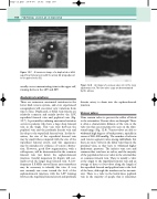
A V
V
Figure 12.7 A transverse image of a duplicated or bifid superficial femoral vein with the artery (A) lying adjacent to the paired veins (V).
usually cross-communicating veins in the upper calf, running between the LSV and SSV.
Anatomical variations
There are numerous anatomical variations in the lower limb venous system, and even experienced sonographers will encounter new variations from time to time. Duplicated, or bifid, vein systems are relatively common and mainly involve the LSV, superficial femoral vein and popliteal vein (Fig. 12.7). A potentially confusing anatomical variation occurs in patients who have a large deep femoral vein in the thigh. This vein runs between the popliteal vein and the profunda femoris vein and lies deep to the superficial femoral vein. In this sit- uation, the size of the superficial femoral vein may be small when compared with the size of the superficial femoral artery, and this appearance may be mistaken for evidence of venous obstruc- tion. However, good flow augmentation, with a calf squeeze, will be demonstrated in the common femoral vein just below the saphenofemoral junction. Careful inspection by duplex will nor- mally reveal the larger deep femoral vein. A low- frequency 3.5 MHz curved linear array transducer may be necessary to identify this vein. A very rare anomaly can occur toward the level of the saphenofemoral junction, with the LSV running between the superficial femoral artery and profunda
An image of a venous valve site in the long saphenous vein. The two valve cusps are demonstrated
by the arrows.
Figure 12.8
femoris artery to drain into the saphenofemoral junction.
Venous valves
Veins contain valves to prevent the reflux of blood to the extremities. Venous valves are bicuspid. There is often a characteristic dilation of the vein at the valve site that can sometimes be seen on the ultra- sound image (Fig. 12.8). Venous valves are able to withstand high degrees of back pressure, typically in excess of 250–300 mmHg. The number of valves in each venous segment varies among individuals, but there are more valves in the distal veins than in the proximal veins, as they have to withstand higher hydrostatic pressures. The inferior vena cava and common iliac vein have no valves, and the majority of the population have no valves in the external iliac or common femoral vein. There is usually a valve at the origin to the superficial femoral vein and an average of three to four valves along the length of the superficial femoral and popliteal vein to the level of the knee, although the number can be inconsis- tent. There is a valve in the below-knee popliteal vein in the majority of people that is sometimes
