
Page 179 - Libro vascular I
P. 179
Chap-12.qxd 29~8~04 14:52 Page 170
170
PERIPHERAL VASCULAR ULTRASOUND
competence of the vein in the upper thigh and at the saphenofemoral junction (Abu-Own et al 1994). Symptoms and conditions caused by varicose veins can include aching, throbbing, burning, venous eczema, bleeding and ulceration. Some patients complain of symptoms that appear out of propor- tion to the size of their veins, which may be only mildly varicose. Some patients with superficial vari- cose veins may also have coexisting deep venous insufficiency.
Treatment of superficial venous disorders
The treatment of abnormal veins varies, depending on the severity of the condition (Browse et al 1999). Thread veins can be treated by injection sclero- therapy, followed by local compression to occlude the vein. They can also be treated by laser. Larger varicose veins can be surgically removed or stripped. In the case of the LSV, the saphenofemoral junction and tributaries are ligated at the groin, and the main trunk stripped with a vein stripper to knee level or below. In the case of the SSV, ligation of the saphenopopliteal junction is performed. Some sur- geons strip the vein, but others leave it intact to avoid injury to the sural nerve, which is closely asso- ciated with the vein. Some surgeons ligate large perforators, and these can be marked preoperatively with the aid of duplex scanning. The remaining veins are then removed or avulsed using small microincisions. It is worthwhile watching some varicose vein surgery as it gives a better appreciation of the anatomy seen during duplex examinations.
A relatively new treatment known as endovenous saphenous obliteration has been introduced with promising results. This technique uses radiofre- quency heating of a catheter tip introduced into the vein, causing collapse of the vein wall. The pro- cedure is carried out under duplex guidance to position the catheters.
Skin changes and venous ulcers
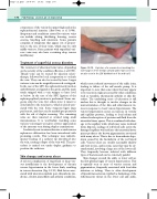
A serious complication of superficial or deep ven- ous insufficiency is the development of chronic venous hypertension in the lower limb, resulting in venous ulceration (Fig. 12.10). Risk factors asso- ciated with ulceration include post-thrombotic syn- drome, obesity, immobility and arthritic conditions,
A picture of a venous ulcer involving the lower aspect of the medial calf and ankle. Varicose veins
are also seen in the LSV distribution of the mid-calf.
Figure 12.10
which cause reduced movement of the ankle joint, leading to failure of the calf muscle pump. It is important to note that some ulcers that may appear to be venous in origin are caused by other conditions, such as vasculitis, rheumatoid arthritis or skin dis- orders. The underlying cause of ulceration is still unclear but is thought to involve changes in the microcirculation of the skin and subcutaneous tis- sues in response to local venous hypertension. The venous hypertension causes an increase in venular and capillary pressure, leading to local edema and reduced reabsorption of proteins and fluid from the interstitial tissue spaces. This is combined with dam- age to the capillary walls, which may cause localized tissue hypoxia. Leakage of red blood cells across the damaged capillary wall and into the interstitial tissue spaces produces the brown pigmentation associated with many ulcers. This is due to hemosiderin depo- sition caused by the breakdown of the red blood cells. Venous ulcers are usually reasonably shallow and vary in size, and in some cases they may be cir- cumferential, involving a large area of the lower calf. They frequently become infected with different types of bacteria and can be extremely painful.
Skin changes around the ankle or lower calf are the first physical signs of venous hypertension. This is typically seen as areas of venous eczema and pigmentation, frequently associated with local skin irritation or itching. There is often development of lipodermatosclerosis, typified as hardening of the subcutaneous tissues in the lower calf and ankle,
