
Page 183 - Libro vascular I
P. 183
Chap-12.qxd 29~8~04 14:52 Page 174
174
PERIPHERAL VASCULAR ULTRASOUND
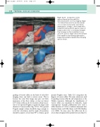
Figure 12.15 Incompetence of the saphenofemoral junction and LSV is demonstrated with color flow imaging. Images 1 to 4 show flow across the junction (arrow) over a 3 s time interval at the end of distal augmentation. In image 1, flow (coded red) is toward the heart just before squeeze release. In images 2 and 3, there is increasing retrograde flow. In image 4, reflux (coded blue) is seen across the junction. Image 5 shows the LSV with flow toward the heart during augmentation. Image 6 demonstrates marked reflux following squeeze release.
12
34
56
grading of venous reflux on the basis of color flow imaging alone can lead to serious errors. This is because the color flow image may give very little impression of the flow volume or may not detect low-volume reflux at all. It is therefore always neces- sary to use spectral Doppler to grade the degree and duration of venous reflux (Fig. 12.16). The spectral Doppler sample volume should be large enough to cover the vein lumen, and the angle of insonation should be equal to or less than 60° to obtain a good
spectral Doppler trace. Table 12.1 categorizes the degrees of venous reflux. The same criteria are used for grading venous reflux by calf compression or the Valsalva maneuver. Although the classification of venous reflux can sometimes be subjective, many contemporary scientific publications have used a reflux duration of 0.5 s to indicate abnormal valve function (Sarin et al 1994, Evans et al 1998, Ruckley et al 2002). In some cases, the reflux may be so severe that retrograde flow persists for more than 4 s.
