
Page 185 - Libro vascular I
P. 185
Chap-12.qxd 29~8~04 14:52 Page 176
176
PERIPHERAL VASCULAR ULTRASOUND
Map 2
150 dB/C 3 Persist Med 2D Opt:Gen Fr Rate:Max
B
SQUEEZE
RELEASE
A
S
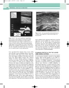
Figure 12.19 Venous stasis (S) is demonstrated as an echogenic speckle pattern in this B-mode image of a deep calf vein.
A 45.4 cm/s B 39.8 cm/s
0.80 cm
There can be problems in quantifying venous reflux. In this example the LSV was very large
(8 mm diameter), but the duration of reflux (0.9 s) is shorter than that shown in Figure 12.16B. However, the volume of reflux is similar to that demonstrated during augmentation. In this example, the volume of blood flow during reflux is probably very significant due to the size of the vein. It should be noted that volume flow calculations are not routinely used in venous examinations.
been speculated that incipient deep venous reflux may occur due to gross superficial varicose veins caus- ing overload of the deep venous system. This may cause some degree of dilation of the deep veins in the lower leg, which impairs normal valve function (Walsh et al 1994). Another problem of interpreta- tion can occur when a vein is very large or dilated, as the duration of reflux may be relatively short. However, the volume of reflux can be high due to the large diameter of the vein. In this situation, significant reflux may be suspected if the shape of the spectral Doppler reflux pattern is the same as the augmenta- tion pattern (Fig. 12.18). It may be difficult to aug- ment flow in the veins of patients with gross venous stasis, and very little reflux will occur following distal release. However, the B-mode image often displays aggregation of the blood in the dilated vein as a speckle pattern (Fig. 12.19). Only a small amount of movement in the speckle pattern will be seen during calf compression. The deep venous sinuses in the calf
Figure 12.18
may be dilated and congested with blood, and this appearance can be mistaken for a deep vein throm- bosis (see Ch. 13). Any problems of interpretation should be reported to the referring clinician, with a print taken of the relevant flow patterns. In some cases, it may be necessary to complement the duplex scan with other diagnostic tests, such as ambulatory venous pressures or plethysmography.
SCANNING PROTOCOL FOR THE LOWER
LIMB VENOUS SYSTEM
A venous preset should be selected on the duplex scanner, which should typically set the PRF at a 1000 Hz. The color wall filter should also be set at a low level. A 10MHz transducer, or broad-band equivalent, is normally used for scanning superficial varicose veins. However, a 5MHz transducer, or broad-band equivalent, is usually required for imag- ing the deep veins and junctions. A combination of B-mode, color flow imaging and spectral Doppler is used throughout the examination. The assessment of venous reflux is performed with the transducer in a longitudinal plane to the vein. However, the assess- ment of perforator competency may require the probe to be orientated into other imaging planes to follow its course. It is necessary to perform an exam- ination of the femoral and popliteal veins during any superficial vein assessment to assess the competency
