
Page 186 - Libro vascular I
P. 186
Chap-12.qxd 29~8~04 14:52 Page 177
ANATOMY OF THE LOWER LIMB VENOUS SYSTEM AND ASSESSMENT OF VENOUS INSUFFICIENCY
177
of these vessels. Also, the superficial veins can act as collateral pathways if the deep veins are obstructed, and surgery of the superficial veins would be con- traindicated and potentially damaging. The tech- nique for assessing varicose veins is described below, but it is often quicker to image the varicose veins distally and follow them back to their supply.
Assessment of the LSV and deep veins
of the thigh and knee
A
V
J
1. The saphenofemoral junction is located by first identifying the common femoral vein in trans- verse section just below the level of the inguinal ligament. The anatomy in this region is demon- strated in Figure 9.6. The common femoral vein lies medial to the common femoral artery and is normally larger than the artery. The transducer is then moved distally, and the saphenofemoral junction will be seen on the medial anterior side of the common femoral vein (Fig. 12.20). It is usual to see other branches dividing from the saphenofemoral junction. Remember that these branches may be the main supply to the varicose veins.
2. The transducer should then be rotated so that the common femoral vein and saphenofemoral junction can be seen in longitudinal section (Fig. 12.21). The competency of the common femoral vein, saphenofemoral junction and proximal LSV can be assessed using distal com- pression and the Valsalva maneuver. The origin of the superficial femoral vein, which lies below the level of the saphenofemoral junction, should also be assessed for competency. Large visible branches dividing from the saphenofemoral junction can also be checked for competency, especially the anterolateral trunk, which can often supply varicose areas in the anterior thigh and calf. Medial branches of anterolateral vein in the upper and mid-thigh region can also supply varicose areas in the medial thigh. They can also run directly into the main trunk of the LSV. In this situation the main proximal trunk of the LSV may be competent.
3. The LSV is examined in transverse section along the medial thigh to the knee. In transverse section, it is often possible to see the LSV running directly into varicose areas. Large perforators, branches
A
L
A
B
V
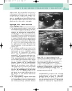
A: A transverse image of the right common femoral vein demonstrating the position of the
saphenofemoral junction (J), common femoral vein (V) and common femoral artery (A). This view is sometimes called ‘Mickey Mouse’ for obvious reasons. In this example, it is likely that the junction is incompetent as the LSV is very large. Note that some small branches are dividing from the junction (arrow). B: A transverse image taken in the upper thigh demonstrating the LSV (L), anterolateral branch (arrow), superficial femoral vein (V) and superficial femoral artery (A).
Figure 12.20
and bifid systems are relatively easy to identify on the transverse B-mode image. Large thigh perforators should be assessed for competency. The LSV is then followed in longitudinal section from the saphenofemoral junction to the knee and assessed for competency at frequent intervals.
