
Page 192 - Libro vascular I
P. 192
Chap-12.qxd 29~8~04 14:52 Page 183
ANATOMY OF THE LOWER LIMB VENOUS SYSTEM AND ASSESSMENT OF VENOUS INSUFFICIENCY
183
Common femoral vein
V
Profunda vein
ALTV
A
B
C
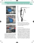
surgery. The femoral vein (V) is seen in all three images. A: The saphenofemoral junction (arrow) has been completely ligated by surgery. B: A small recurrent junction (arrow) is seen to supply a diffuse network of small veins. C: The SFJ has been ligated but a diffuse web of small veins are seen adjacent to the vein (arrow).
SFV
V
LSV
Varicose veins
Incomplete ligation of the sapheno- femoral junction can be the cause of recurrent varicose
Figure 12.30
veins. In this example the anterolateral thigh vein (ALTV) has been left intact following incomplete ligation of the junction (arrow). The ALTV now supplies the varicose areas shown on the diagram.
Incompetent tributaries
In some cases, the saphenofemoral junction has been ligated, but small tributaries from the inner aspect of the groin supply varicose areas in the LSV distribution or the main LSV trunk, if it has not been stripped. These tributaries are often supplied from pudendal and perineal veins. Typically, the scan will demonstrate varicose veins, or an intact trunk if this has not been stripped in the upper thigh, breaking up into small tributaries before disappearing toward the inner aspect of the groin. These tributaries are usually seen as a fine, diffuse web of veins on the color flow image. Sometimes these tributaries may run close to the femoral vein, but no direct connection will be identified (Fig. 12.29C). It is also possible for recurrent varicose
Three examples of ultrasound findings at the level of the saphenofemoral junction following
Figure 12.29
V
