
Page 191 - Libro vascular I
P. 191
Chap-12.qxd 29~8~04 14:52 Page 182
182
PERIPHERAL VASCULAR ULTRASOUND
A
SV
DV
P
INVESTIGATION OF RECURRENT
VARICOSE VEINS
Some patients develop secondary or recurrent varicose veins over a variable time period following surgery. The scanning technique for the investiga- tion of recurrent varicose veins is very similar to that used for the investigation of primary varicose veins. However, it is important to keep an open mind as to the source of the recurrent veins, as their supply can be unpredictable. It is often easier to begin the examination at the level of the vari- cose areas in transverse section and work proxi- mally to the point of supply. The use of color flow imaging during calf augmentation can allow smaller varicose veins to be followed proximally if the B-mode imaging is poor. Some of the main causes of recurrent varicose veins are summarized below. However, it may be some considerable time before the trainee sonographer is familiar with all the variations.
Possible causes of LSV recurrences
Incomplete ligation of the saphenofemoral junction
Normally the junction should be ligated and any tributaries divided (Fig. 12.29A). However, due to misidentification or inadequate dissection, it is possible to ligate only a small tributary during sur- gery, rather than the main junction. The level of the saphenofemoral junction should be examined in transverse section, where it is easy to identify a large patent junction. Sometimes the scan demonstrates that the main trunk of the LSV has been ligated just distal to the saphenofemoral junction but a tributary has been left intact. This tributary then supplies the varicose veins or intact LSV trunk, if it has not been stripped. This is often the case when the anterolateral saphenous vein is found to be intact at the level of the saphenofemoral junction. It frequently supplies varicose areas in the anterior and medial aspects of the thigh, which in turn run into the calf (Fig. 12.30). Occasionally, there may be a very small recurrent junction that can be difficult to identify without the aid of color flow imaging (Fig. 12.29B).
VV
B
P
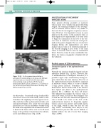
A: A venogram demonstrating a perforator (P) running between the deep veins (DV) and
superficial varicose veins (V V). B: A B-mode image demonstrating a large perforator (P). The fascia is demonstrated by the arrows, and there is a clear break in the fascia as the perforator runs between the deep vein (DV) and superficial vein (SV).
the skin surface. Occasionally a large localized dila- tion can be seen in the main trunk, called a varix. Sometimes the supplying vein may appear reason- ably small, but reflux is demonstrated with color and spectral Doppler. The easiest way of locating perforators is to run the transducer steadily along the trunk of the superficial vein in transverse section. A break in the fascia will be seen on the B-mode image as the perforator runs between the subcuta- neous and subfascial areas (Fig. 12.28).
Figure 12.28
DV
