
Page 193 - Libro vascular I
P. 193
Chap-12.qxd 29~8~04 14:52 Page 184
184
PERIPHERAL VASCULAR ULTRASOUND
directly, or via side branches, to the perforator. Perforators to the deep veins can have very tortu- ous courses. If a mid-thigh perforator is the main supply to the varicose areas, there may be only small, or no, varicose veins seen above the level of the perforator. Duplex scanning can be used to mark the location of perforators preoperatively.
Incomplete stripping of the LSV trunk in the thigh
Sometimes the saphenofemoral junction has been ligated, but the vein stripper has been passed down a bifid trunk of the LSV or a branch, leaving the majority of the main trunk intact. The image will demonstrate the presence of an incompetent LSV trunk that breaks into small tributaries toward the groin with a variable supply, as described in some of the examples above.
Secondary varicose veins
Over time, it is not uncommon for secondary vari- cose veins to develop in the thigh and calf that were not visible when the original surgery was per- formed. These are often imaged as numerous tor- tuous varicose veins that may lie very superficially in the LSV distribution. They often have a diffuse supply, sometimes involving superficial tributaries in the groin.
Incompetence of the SSV
Varicose veins in the LSV distribution of the calf can occur due to incompetence of the SSV. In this situation, posteromedial varicose branches of the SSV interconnect to the LSV distribution (Fig. 12.22B).
Possible causes of SSV recurrences
Incomplete ligation of the saphenopopliteal junction
Recurrence can occur if there has been incomplete ligation or misidentification of the saphenopo- pliteal junction during surgery. With the transducer in transverse section, the SSV can be followed
Recurrent saphenofemoral junction
LSV
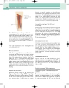
Figure 12.31 A diagrammatic representation of neovascularization. There is a small recurrent connection between the femoral vein and a diffuse network of veins in the groin and upper thigh. In this example the main trunk of the LSV has not been stripped and is supplied by the small diffuse veins. The ultrasound appearance at the level of the femoral vein will be similar to that shown in Figure 12.29B.
veins to be supplied from veins running from the lower abdominal wall.
Neovascularization
It has been suggested that neovascularization or ‘re-growth’ can occur between the femoral vein and superficial varicose veins at the groin following liga- tion of the saphenofemoral junction (Jones et al 1996) (Fig. 12.31). These small veins then supply the LSV, if it had not been stripped, or proximal sec- ondary varicose veins. Duplex scanning will reveal a diffuse web of small tortuous veins in the groin, with a small connection to the femoral vein that may be impossible to follow without the aid of color flow imaging.
Thigh or calf perforators
Recurrent varicose veins can be supplied by thigh or calf perforators (Fig. 12.28). Large thigh perforators are relatively easy to identify by follow- ing the varicose trunk up the medial thigh in trans- verse section, where it will be seen to connect
