
Page 195 - Libro vascular I
P. 195
Chap-12.qxd 29~8~04 14:52 Page 186
186
PERIPHERAL VASCULAR ULTRASOUND
these circumstances it is very difficult to examine the function of the veins, but it may be an indication that the leg is infected. Appropriate action, such as antibiotic therapy and leg elevation, may need to be taken to reduce the infection or cellulitis. The leg can be reassessed when the hyperemia subsides.
OTHER DISORDERS OF THE VENOUS
SYSTEM
Superficial thrombophlebitis
Superficial thrombophlebitis is an inflammatory process that involves the superficial veins (see Ch. 13). The superficial vein may become partially or fully thrombosed. Typically, the area around the phlebitis is reddened, tender and hot, and the super- ficial vein may be swollen and hard. Phlebitis is nor- mally treated with analgesia and anti-inflammatory drugs, but superficial vein stripping may be required, especially if there is a thrombus tip extending to the saphenofemoral junction or saphenopopliteal junction.
Klippel-Trenaunay syndrome (KTS)
Klippel-Trenaunay syndrome is a congenital con- dition and consists of a range of abnormalities
in the lower leg. The arterial signal, above the baseline, demonstrates high-volume flow throughout the cardiac cycle. There is continuous high-volume flow in the vein, shown below the baseline.
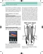
Vein scan report
that can involve the skin capillaries, often causing nevi (birthmarks or port wine stains), bone and soft tissue hypertrophy (excessive limb growth) and venous varicosities. Each case of KTS is unique, and often only one limb is affected, but other areas of the body may also be involved. Abnormalities of the venous system range in severity (Browse et al 1999). Visible varicose veins vary from very minor to severe and can be widely distributed throughout the leg. Varicosities are commonly seen on the lateral aspect of the thigh and calf. In some cases of KTS, the deep veins may be abnormal. Abnormalities can include absence of parts of the deep venous system, unusu- ally small deep veins or large, dilated deep veins with nonfunctioning valves. It is therefore very important to scan the deep venous system in all patients with
Right
Left
LSV normal
LSV reflux
Deep venous reflux
SSV reflux
Deep veins normal
SSV normal
An example of hyperemic flow patterns in the superficial femoral artery and vein due to infection
Figure 12.32
Patient details
The use of diagrams makes it easier for the clinician to interpret the findings of a venous duplex
examination (see text).
Figure 12.33
