
Page 189 - Libro vascular I
P. 189
Chap-12.qxd 29~8~04 14:52 Page 180
180
PERIPHERAL VASCULAR ULTRASOUND
and proximal SSV can be extremely tortuous, with the vein doubling back on itself in an S-shape pattern longitudinally, while following a tortuous path in either the medial or lateral direction. This leads to a very confusing image in which it is even possible to see different sec- tions of the proximal SSV in the same scan plane. Slow, careful movement of the probe should be used to track the vein back to the popliteal vein. It is also possible to mistake branches of the gastrocnemius vein for the SSV if they run superficially within the gastrocne- mius muscle. Care should be used when identi- fying the anatomy in this area. Finally, large perforators, separate from the SSV, can some- times be found in the popliteal fossa, supplying superficial varicose areas.
2. The saphenopopliteal junction and proximal SSV are then imaged in longitudinal section (Fig. 12.25). In some cases, the junction is
tortuous, and rotation of the probe is required to allow the junction to be visualized. The saphenopopliteal junction and proximal SSV should then be assessed for reflux with a strong calf squeeze. If not already performed previously, the competency of the above- and below-knee popliteal vein should be assessed.
3. Unlike the saphenofemoral junction, there is considerable anatomical variation in the position of the saphenopopliteal junction (Figs 12.6 and 12.26). In some cases it may be impossible to identify, owing to the height of the junction in the posterior thigh. The Giacomini vein, if pres- ent, can be the source of SSV incompetence. In this situation, it is possible to follow the Giacomini vein, which will be seen to run as a continuation of the SSV, in transverse section up the posterior thigh to its origin, which may be from a number of sources. These include the LSV, posterior thigh perforator or veins at the top of the posterior thigh. In the latter case, it may be impossible to identify the source, which
SSV
J
PA
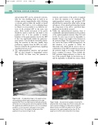
proximal SSV. There is a small deep vein (arrow) joining the SSV at the level of the junction (J) to the popliteal vein (PV). The popliteal artery (PA) lies below the vein. It is not always possible to see the junction in this plane or this clearly, especially if it lies to the medial or lateral side of the popliteal vein as shown in Figure 12.24.
GS GV
J
PV
up the posterior thigh as the Giacomini vein (G). A gastrocnemius vein (GV) also drains to the SSV just proximal to the saphenopopliteal junction (J). The popliteal vein (PV) is demonstrated in this image.
PV
A longitudinal image of the popliteal fossa demonstrating a dilated saphenopopliteal junction and
Figure 12.25
An anatomical variation involving the proximal SSV. In this image the SSV (S) continued to run
Figure 12.26
