
Page 199 - Libro vascular I
P. 199
Chap-13.qxd 1~9~04 16:43 Page 190
190
PERIPHERAL VASCULAR ULTRASOUND
incidence was highly dependent upon age and increased from 2–3 per 10 000 person years at age 30–49 years to 20 per 10000 person years at age 70–79 years. Around 40% cases of DVT were found to be idiopathic (of unknown cause). The annual rate of PE is somewhere in the region of 6 cases per 10000 in the general population (Nicolaides et al 1994). A review by White (2003) indicated that death occurs in approximately 12% of PE cases within 1 month of diagnosis. Open or healed venous ulceration occurs in around 1% of the general adult population, with a proportion attributed to post- thrombotic syndrome (Fowkes et al 2001). The early detection and treatment of DVT can therefore reduce the subsequent risk of mortality or long-term morbidity.
Virchow (1846) described the association between thrombosis in the legs and emboli in the lung. The factors predisposing to thrombosis are described by his famous triad of coagulability of the blood, damage to the vein wall or endothelium and venous stasis. Venous thrombi are believed to originate in valve cusp pockets (Fig. 13.1) or in the deep venous muscular sinuses, such as the soleal veins. DVT most commonly occurs in the calf veins and can propa- gate to the proximal veins. It is not necessary for all the calf veins to be affected in order for proximal propagation to occur. It is believed that approxi- mately 10–20% of calf vein thrombi propagate to
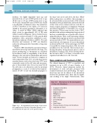
Figure 13.1 A longitudinal B-mode image of the below- knee popliteal vein. An area of thrombus (arrow) is seen to extend from the valve site on the posterior wall.
the deep veins across and above the knee (Khaw 2002, Labropoulos et al 2002). This is thought to be associated with an increased risk of PE. Isolated thrombosis of the proximal veins, such as the femoral or iliac veins, is less common and can occur due to trauma, surgery, pregnancy or malignancy. Proximal and distal propagation of thrombus can occur in this situation. Venous stasis can also lead to DVT, and this is why patients undergoing long periods of bed rest or immobility are at greater risk of devel- oping thrombosis. There is also increasing evidence to suggest that long-haul air travel is associated with an increased risk of DVT. The main risk factors asso- ciated with the development of venous thrombosis are shown in Box 13.1. In the early stages of a DVT, it is possible for a large proportion of the clot to be nonadherent to the vein wall. This is termed a free- floating thrombus. In this situation there is a risk of detachment, leading to PE. As free-floating throm- bus becomes older (7–10 days), it becomes more organized and adherent to the vein wall.
Signs, symptoms and treatment of DVT
The clinical diagnosis of DVT is unreliable and inaccurate in up to 50% of cases (Cranley et al 1976). However, typical symptoms include the development of acute calf pain associated with localized tenderness, heat and swelling. The super- ficial veins may also be dilated. If the thrombosis involves the proximal veins, there may be signifi- cant swelling of the thigh. Unfortunately, other
Box 13.1 Risk factors for the development of deep vein thrombosis
● Coagulation disorders
● Immobilization
● Surgery and trauma
● Malignancy
● Septicemia
● Oral contraceptives
● Increasing age
● Stroke
● Heart failure
● Previous history of deep vein thrombosis
● Long-haul air travel
