
Page 200 - Libro vascular I
P. 200
Chap-13.qxd 1~9~04 16:43 Page 191
DUPLEX ASSESSMENT OF DEEP VENOUS THROMBOSIS AND UPPER LIMB VENOUS DISORDERS
conditions, such as cellulitis and edema, can mimic the symptoms of DVT. In some cases of DVT the patient may be asymptomatic, especially if the throm- bus is small. In extreme cases of DVT the outflow of the limb is so severely reduced that the arterial inflow may become obstructed, leading to venous gangrene. This condition is called phlegmasia cerulea dolens. The foot may appear blackened and the limb swollen and blue, even when elevated.
PE occurs when a segment of clot breaks loose, travels through the right side of the heart and lodges in branches of the pulmonary artery. This leads to a perfusion defect in the arterial bed of the lungs. The symptoms of PE include the following:
● sudden breathlessness
● pleuritic chest pain
● coughing up of blood
● right-sided heart failure or cardiovascular collapse
● death.
Radioisotope studies are frequently used for investi- gating perfusion and ventilation defects in the lungs. The prevention of DVT includes the use of elastic support stockings, which increase venous return and therefore reduce the risk of venous stasis. Patients at high risk may be advised to take aspirin or be given low molecular weight heparin if they are undertaking any activity or treatment that may increase the risk of DVT. Treatment of DVT is usually with antico- agulation drugs. The initial treatment is by an intra- venous infusion of heparin, which is then converted to long-term therapy with oral anticoagulants, such as warfarin. Occasionally, devices called vena caval filters are positioned in the vena cava to catch clots, when there is a high risk of embolization to the lungs. Surgery is sometimes performed to remove
thrombus from the femoral and iliac veins.
The investigation and treatment of isolated calf vein thrombosis remain a contentious issue (Lohr et al 1991, Meissner et al 1997). It is beyond the scope of this book to consider the debate in any detail, but sonographers should be aware of the controversies surrounding this area. Some clinicians will always investigate and treat calf DVT, whereas others will not specifically ask for the calf veins to be examined. Some units perform serial scans of the popliteal vein over a period of 3–5 days to identify any propagation of calf vein thrombi to the
popliteal vein.
Image not available
191
Investigations for diagnosing DVT
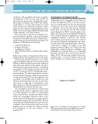
Traditionally, x-ray venography was the main test used for the diagnosis of DVT. It involves an injec- tion of a contrast agent into the venous system via a dorsal foot vein. In some cases it proves impossible to cannulate a foot vein, and in some situations patent deep calf veins do not fill with contrast agent (Bjorgell et al 2000). Currently, duplex scan- ning is the main method of imaging DVT, but it is often combined with pre-imaging tests, in a defined management pathway or protocol. The develop- ment of these protocols has occurred as a result of the increased workload and financial costs experi- enced by most ultrasound departments. These pro- tocols may be complex; an example of one such pathway is shown in Figure 13.2. The process often begins with a clinical assessment, including a risk probability score derived from a set of standard questions. The lower the score, the lower the prob- ability of DVT. The next stage usually involves a bio- chemical assay to measure D-dimer levels in the blood. D-dimers are products that are formed by
An example of a screening protocol for DVT. RPA, risk probability assessment; LMWH, low molecular-
weight heparin. (After Khaw 2002, with permission.)
Figure 13.2
