
Page 202 - Libro vascular I
P. 202
Chap-13.qxd 1~9~04 16:43 Page 193
DUPLEX ASSESSMENT OF DEEP VENOUS THROMBOSIS AND UPPER LIMB VENOUS DISORDERS
193
there is thrombus in the vein it will not collapse. This technique is demonstrated in Figures 13.3 and 13.4. It should be noted that fresh thrombus, which is soft, can partially deform. Compression should be applied at frequent intervals along the length of a vein to confirm patency. Partial collapse of the vein suggests the presence of nonoccluding thrombus. In this situation, the adjacent artery may be seen to deform as the probe pressure is increased to confirm partial obstruction in the vein.
Transducer compression should be applied in the transverse imaging plane rather than the longitudi- nal plane. This is because it is easy to slip to one side of the vein as pressure is applied in the longi- tudinal plane, and this may mimic compression of the vein when observed on the B-mode image. Unfortunately, in some areas the veins lie too deep for compression to be used, such as in the pelvis and sometimes at the adductor canal or calf. Color flow imaging is useful for demonstrating patency in this situation.
The following guidelines can be used in any sequence, depending upon the areas that require assessing. It is sometimes easier to locate a specific vein by looking for the adjacent artery, especially in the calf. The reader should also refer to Chapter 9 for more details on the probe positions for imaging the calf vessels and the main vessels in the thigh and pelvis.
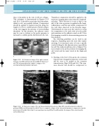
1. Starting at the level of the groin, the common femoral vein is imaged in transverse section and will be seen to lie medial to the common femoral artery (Figs 13.3A and 9.6). The com- mon femoral vein should be compressed to
CFA
CFA
CFV
AB
Figure 13.3 A: A transverse image of the right common femoral vein (CFV) and femoral artery (CFA). B: Patency of the CFV is demonstrated by complete collapse of the
vein (arrow) during transducer pressure.
A
V
A
V
PT
V
PER
A
Figure 13.4
VA AB
A: Transverse image of the calf demonstrating the posterior tibial (PT) veins (V) and arteries (A) and peroneal (PER) veins (V) and arteries (A). B: There is complete collapse of the veins with transducer compression, but the arteries are still visible. Note that it can sometimes be very difficult to differentiate the image of the veins from the surrounding tissue.
